
Diagnosis: Verrucous haemangioma
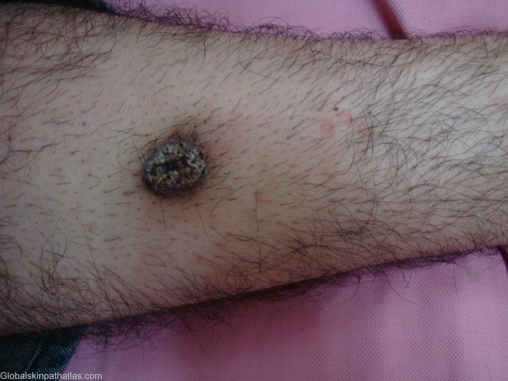
Description: Painless, dark-blue, warty nodule on the leg.
Morphology: Nodule,black
Site: Leg
Sex: M
Age: 22
Type: Clinical
Submitted By: Nameer Al-Sudany
Differential Diagnosis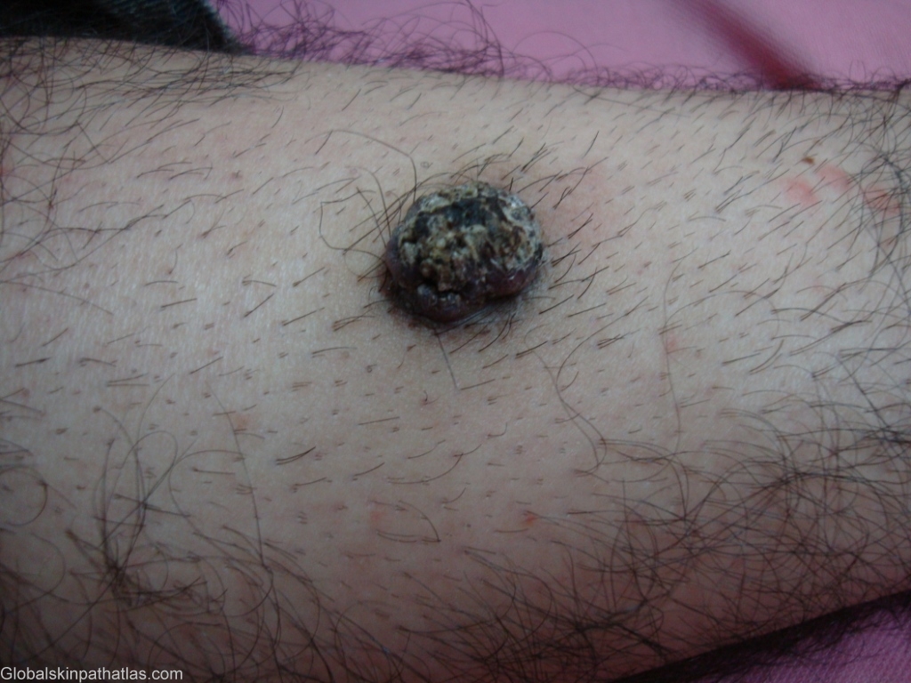
History:
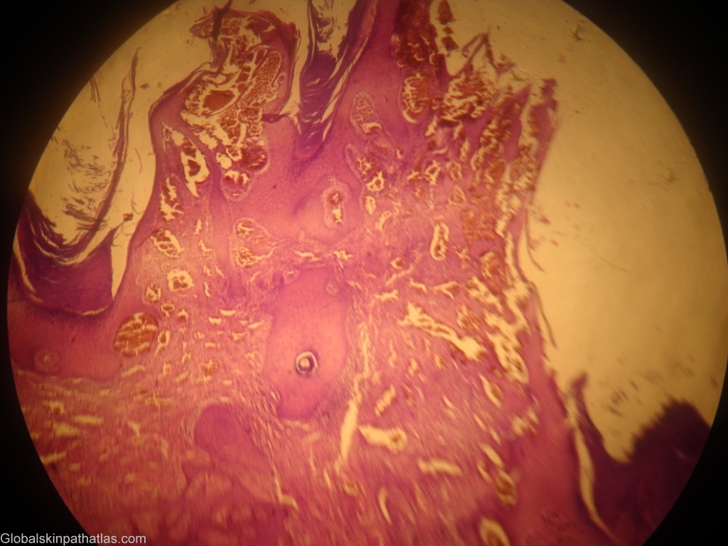
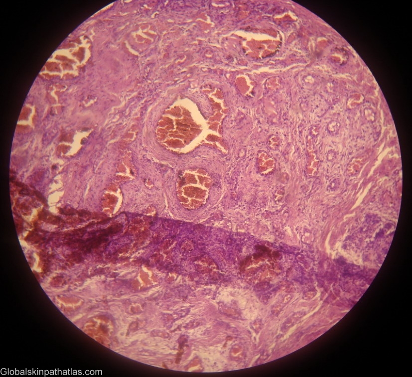
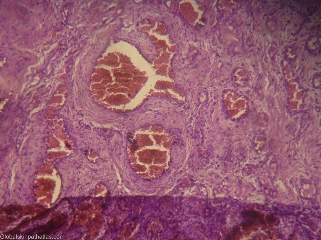
A 22-year-old man gave a history of flat, reddish-blue solitary lesion on his leg since early childhood. At last one year the lesion started to rise above the level of the skin and acquired rough surface. On examination, the lesion was painless, wart-like, dark blue nodule about 2-3 cm in diameter with dome-shaped verrucous surface on the mid of anterior aspect of the leg. Lesional biopsy showed hyperkeratosis, and irregular acanthosis overlying a hemangioma composed of large vascular channels lined by flattened endothelial cells and are surrounded by a layer of pericytes. Verrucous hemangioma is a rare vascular malformation almost invariably present at birth, but may appear later, even in adult life. It presents as well-defined, dark-red, macular areas resembling port-wine stains. After a variable number of years, lesions start to take on their characteristic bluish black hue and an increasingly verrucous surface. The presence of small satellites lesions is rather typical. Recurrent bleeding and infection often cause the patient to seek medical advice for the first time at this stage.