
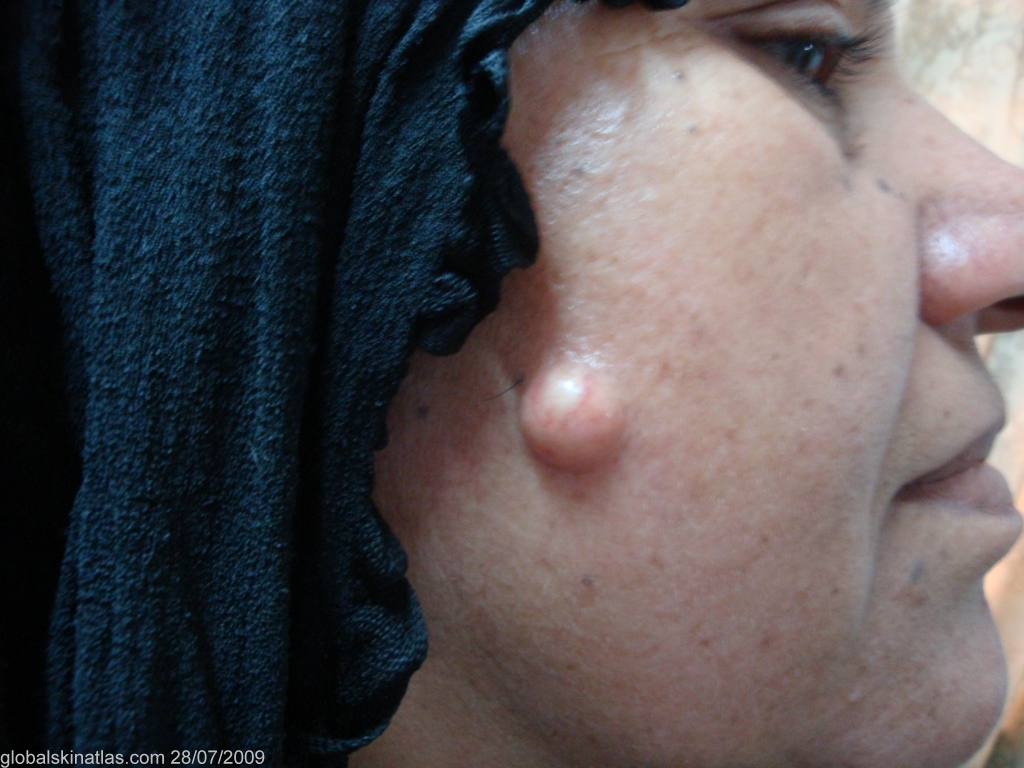
Diagnosis: Epidermoid cysts
Description: Asymptomatic, skin-colourd nodule.
Morphology: Nodule
Site: Cheek
Sex: F
Age: 48
Type: Clinical
Submitted By: Nameer Al-Sudany
Differential DiagnosisHistory:
A dome-shaped, skin-coloured, about 2 cm diameter, asymptomatic nodule with surface telangiectasia in the center of the right cheek of more than one year duration. On clinical background, the main two differential diagnoses were epidermoid cyst and giant mulloscum contagiosum. The lesion has been removed in toto under local anesthesia and found to be an epidermoid cyst.