
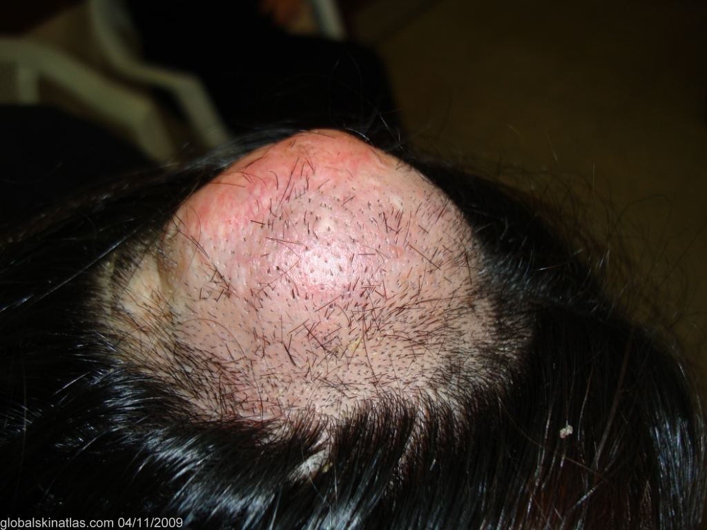
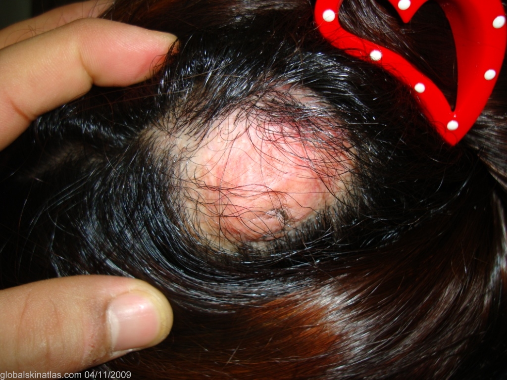
Diagnosis: Cutaneous meningioma
Description: Hard tumour.
Morphology: Nodule
Site: Scalp
Sex: F
Age: 28
Type: Clinical
Submitted By: Nameer Al-Sudany
Differential Diagnosis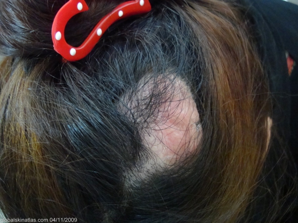
History:
This lady presented with a very hard, painless, large nodular mass on the scalp. It has been noticed since first year of life and increased in size gradually. A skin biopsy was taken and showed an epidermal atrophy, superficial dermal scarring and a tumor infiltrating the reticular dermis and subcutis. The tumor consisted of strands, sheets and nests of uniform cells with large oval vesicular nuclei and granular cytoplasm with frequent cellular whorls, a picture consistent with cutaneous meningioma.
Primary cutaneous meningioma is a developmental defect resulting from the presence of meningocytes outside the calvarium. Small, hard, fibrous, calcified nodules occur along the spine, in the scalp, on the forehead, or rarely in the external ear canal. Most occur on the scalp, some have an underlying connection to the CNS or an underlying bony abnormality, and usually come to medical attention in the first year of life. Diagnosis is made by histologic examination.