
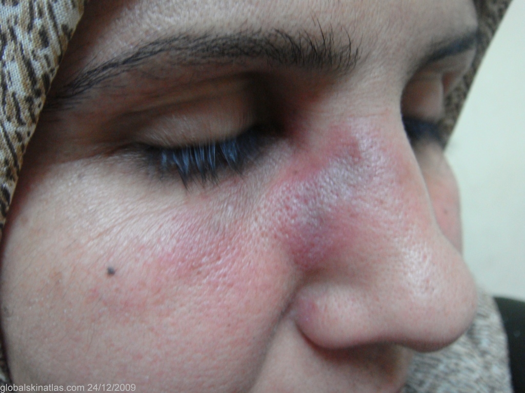
Diagnosis: Discoid lupus erythematosus
Description: Dull red swollen plaques.
Morphology: Plaque
Site: Face
Sex: F
Age: 34
Type: Clinical
Submitted By: Nameer Al-Sudany
Differential Diagnosis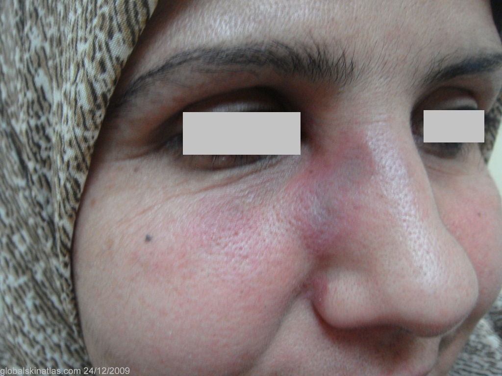
History: DLE typically is characterized by dull-red macules and patches with adherent scales extending into putulous follicles (Carpet's tack scales. The patches usually heal centrally first, with atrophy, scarring, dyspigmentation and telangectasia. However, some discoid lesions are bright red or even urticarial that may mimic drug eruption, contact dermatitis, polymorphous light eruption or even EM. The lesions on the face of the presented case were dull red swollen plaques of about 6 months duration. The ESR was high and the ANF was positive, however skin biopsy was denied by the patient.