
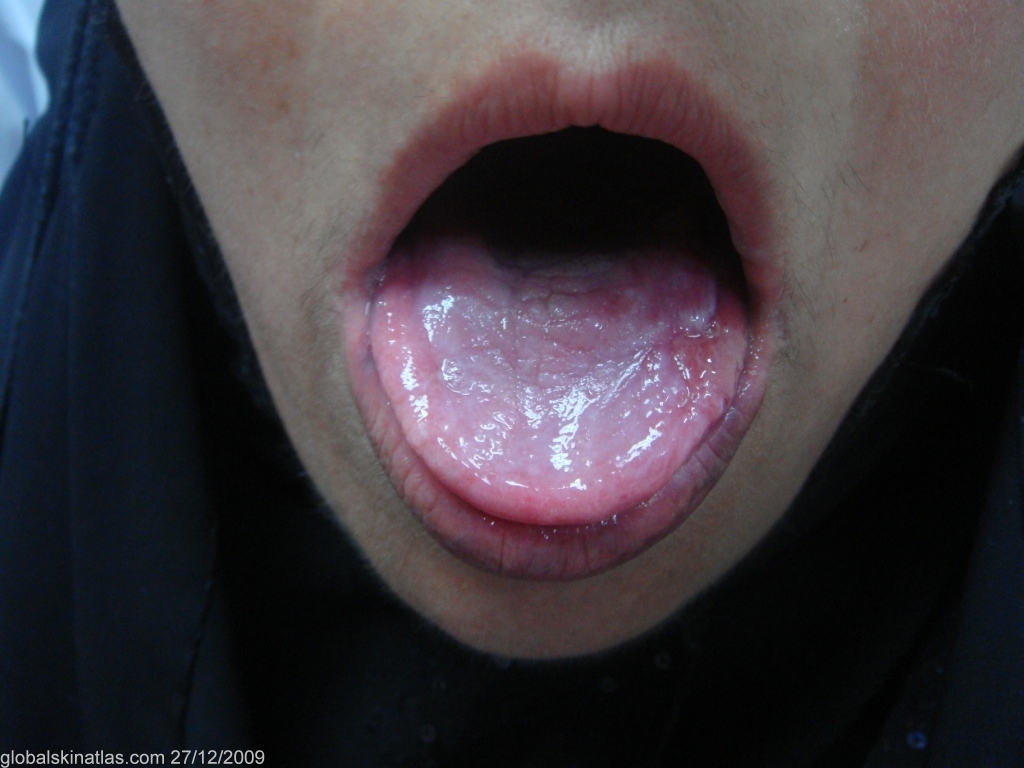
Diagnosis: Lichen planus oral
Description: Erythematous atrophic oral lichen planus.
Morphology: Atrophy
Site: Tongue
Sex: F
Age: 25
Type: Clinical
Submitted By: Nameer Al-Sudany
Differential DiagnosisHistory: Oral lichen planus is a stable but chronic condition as spontaneous remission in an average 5-year follow-up is < 3%. Most cases (75%) occur in women. Oral lesions may be ulcerative (erosive), erythematous (atrophic) or reticular. Patients may simultaneously have several patterns as reticulate lesions are almost always also seen in patients with erythematous type. Symptoms are least common in patients with the “classic” reticulate lesions (only when the tongue is involved). All patients with erosive lesions are symptomatic. The buccal mucosa is involved in 90% of cases, and the gingiva in more than 50%. On the tongue and palate lesions are often mistaken for leukoplakia. The present woman has had an erythematous atrophic variant of oral lichen planus presented as an asymptomatic white glistening and atrophic dorsal surface of the tongue of more than one year duration.