
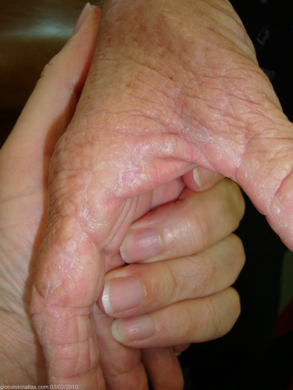
Diagnosis: Collagenous and elastotic marginal plaques
Description: Hyperkeratotic indurated skin of first web space
Morphology: Keratoderma
Site: Web spaces
Sex: F
Age: 60
Type: Clinical
Submitted By: Ian McColl
Differential Diagnosis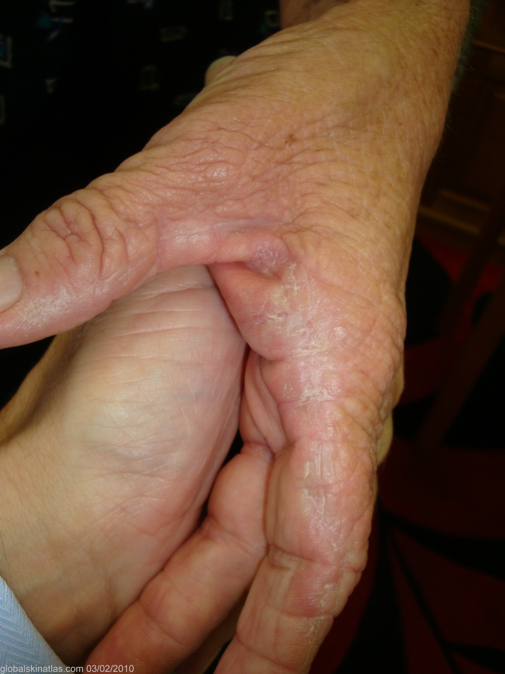
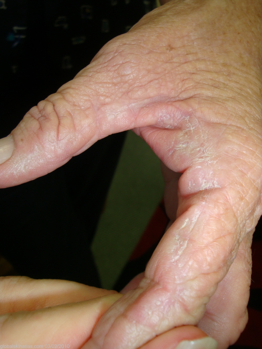
History: A lady in her 60s with thickened hyperkeratotic skin only in the first web spaces of both hands. She had had a lot of sun exposure but denied excessive physical work with her hands. This condition is called Collagenous and elastotic marginal plaques of the hands. The following is from a paper in the AJD Australas J Dermatol. 2001 Aug;42(3):211-3 by Mortimer and Conrad on the condition. " A skin biopsy showed an acellular zone in the reticular dermis composed of thickened bundles of collagen haphazardly arranged, some perpendicular to the epidermis, admixed with elastic fibres and amorphous basophilic elastotic material. Granular calcium deposits were identified, particularly within degenerate collagen bundles. These clinical and histological features are diagnostic of collagenous and elastotic marginal plaques of the hands, a slowly progressive but largely asymptomatic condition. Actinic degeneration and chronic pressure have been proposed as aetiological agents."