
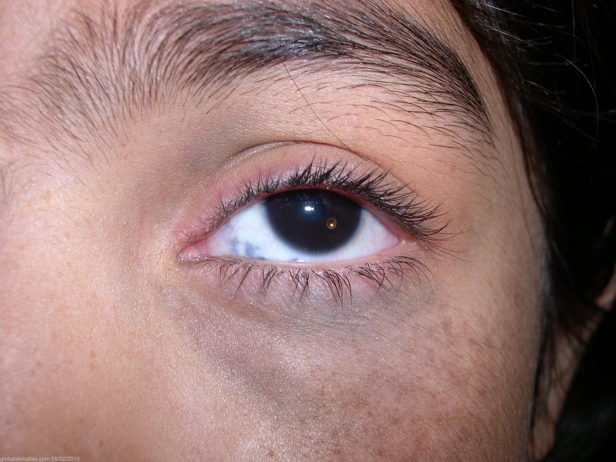
Diagnosis: Nevus of Ota
Description: Bluish grey macules
Morphology: Hyperpigmentation
Site: Cheek
Sex: F
Age: 25
Type: Clinical
Submitted By: Shahbaz Janjua
Differential Diagnosis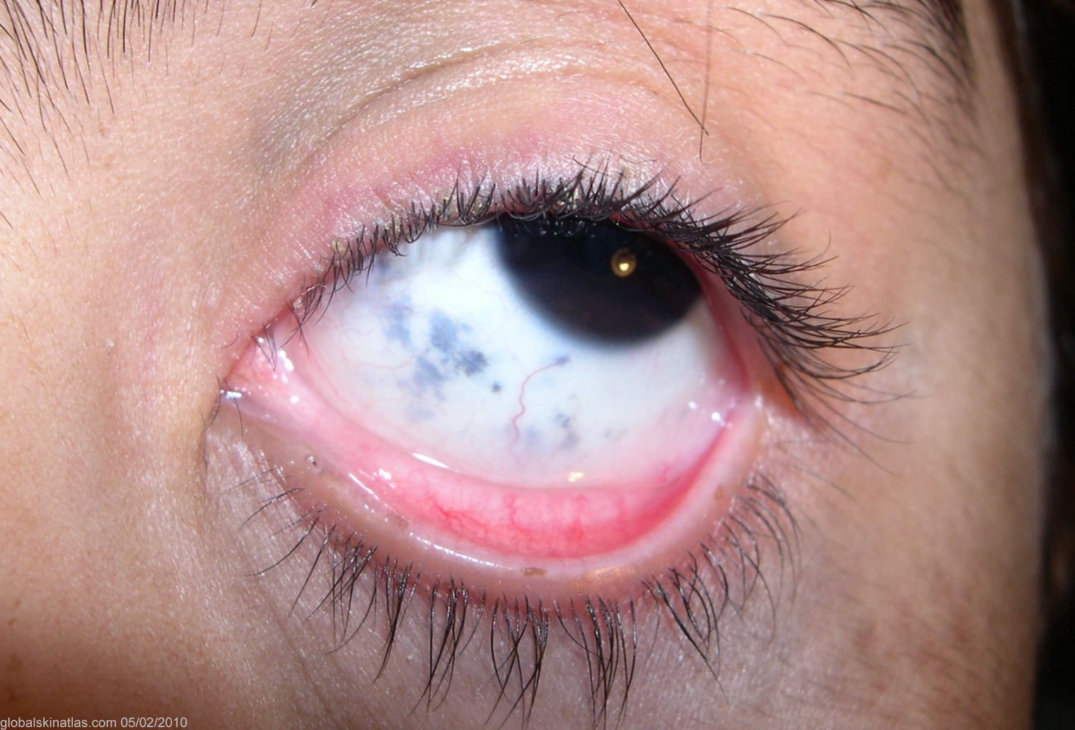
History: Nevus of Ota was described by Ota and Tanino in 1939. Clinically, it presents as blue-to-gray speckled or mottled coalescing macules or patches affecting the forehead, temple, malar area, or periorbital skin. within the distribution of the ophthalmic and maxillary branches of the trigeminal nerve. In addition to skin, pigmentation of nevus of Ota may involve oral mucosa and ocular structures such as the sclera, retrobulbar fat, cornea, and retina.