
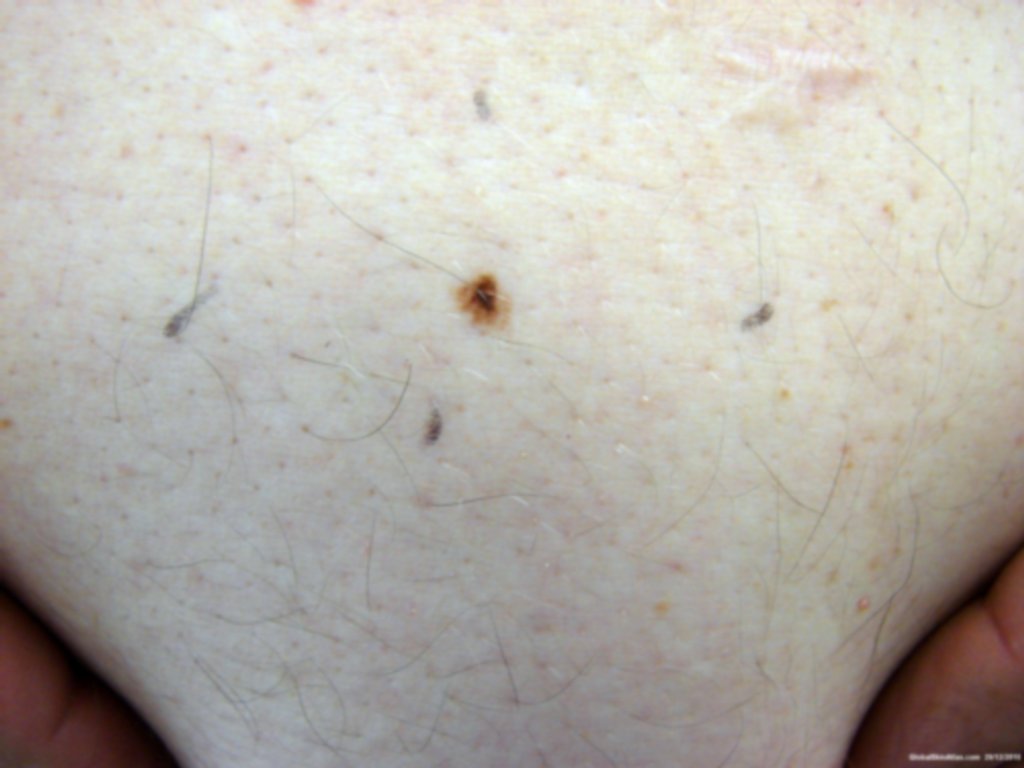
Diagnosis: Melanoma
Description: Pigmented macule
Morphology: Macule brown
Site: Back
Sex: M
Age: 42
Type: Clinical
Submitted By: Ted Rosen
Differential Diagnosis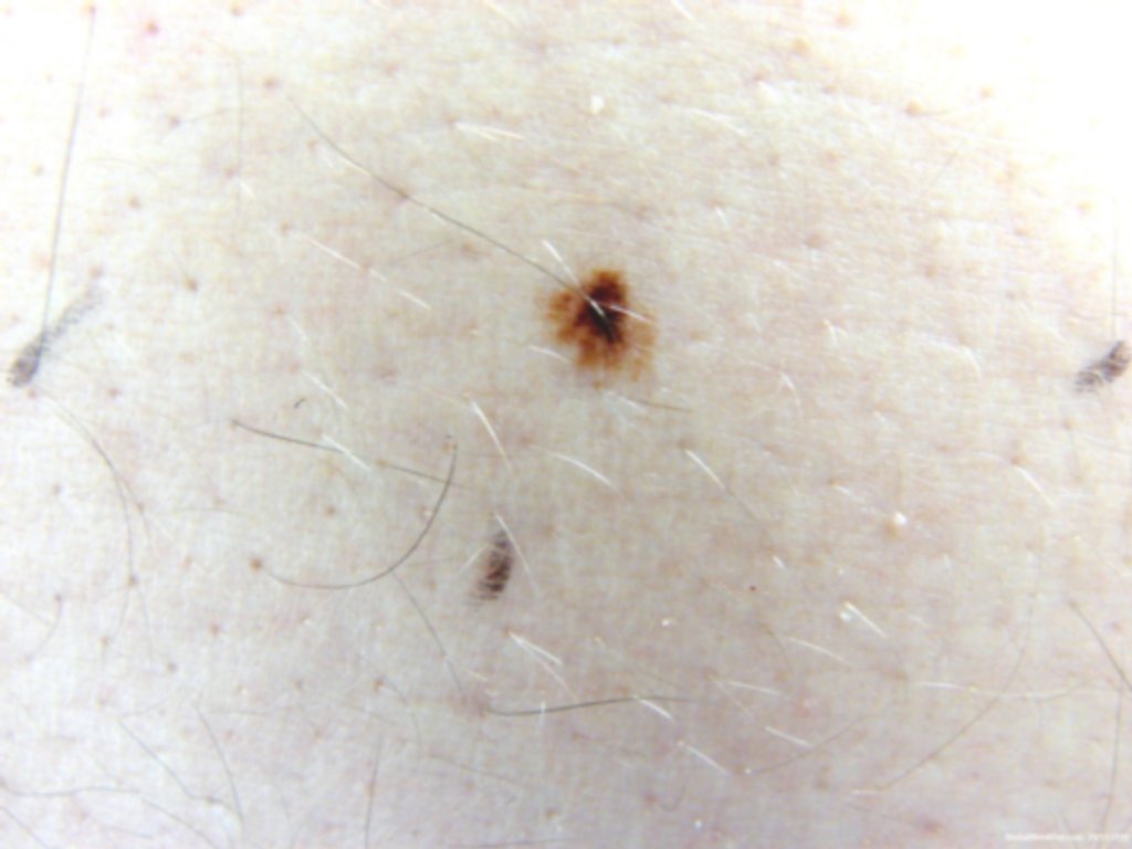
History:
42 year-old referral from primary care physican due to "funny looking" lesion on back. Exam shows clear multiple colors and asymmetry, suggested of melanoma. Lesion excised. 2 of 3 pathologists called it melanoma in situ; one pathologist called it nevus woth severe cytologic atypia and architectural disorder. This highlights the difficulty in pathological interpretation of pigmented lesions. Best to err on the conservative side: assume the worst, as I did with this lesion.