
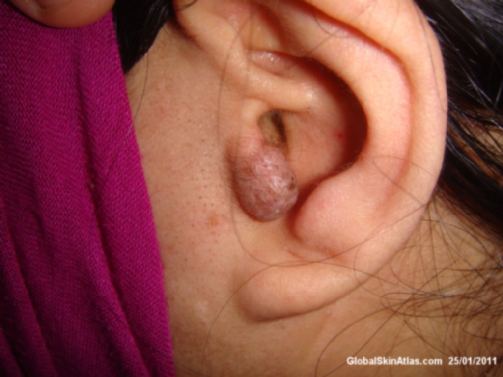
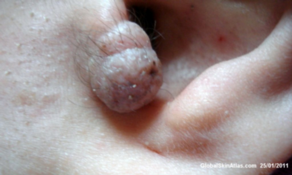
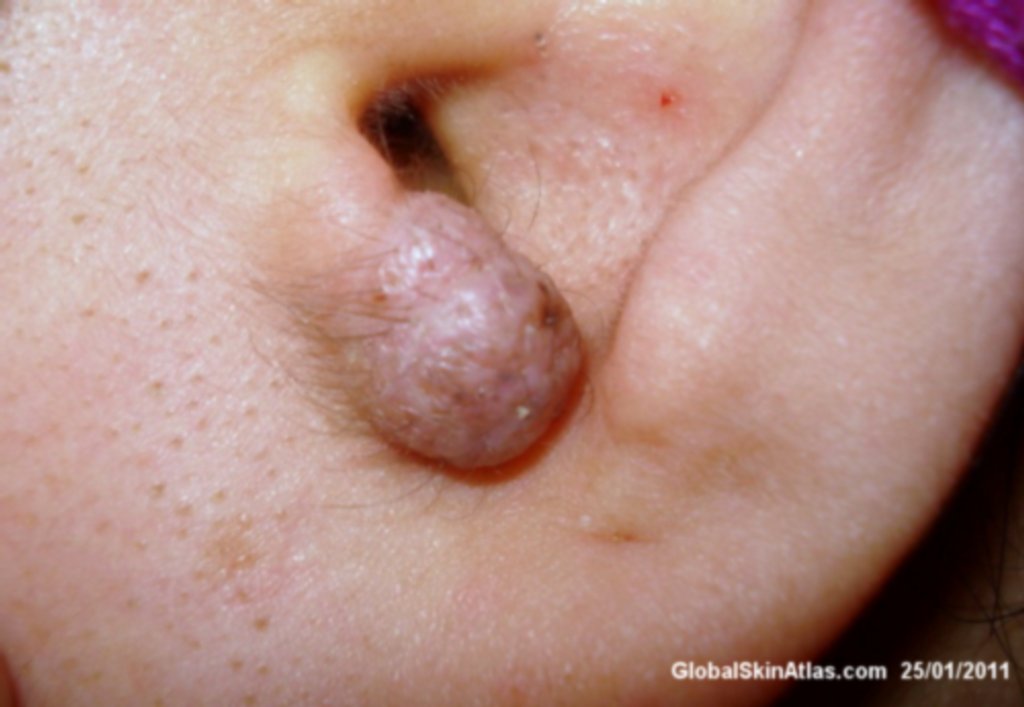
Diagnosis: Angiolymphoid Hyperplasia with Eosinophilia
Description: Solitary pinkish preauricular nodule.
Morphology: Nodule pink
Site: Ear
Sex: F
Age: 30
Type: Clinical
Submitted By: Nameer Al-Sudany
Differential Diagnosis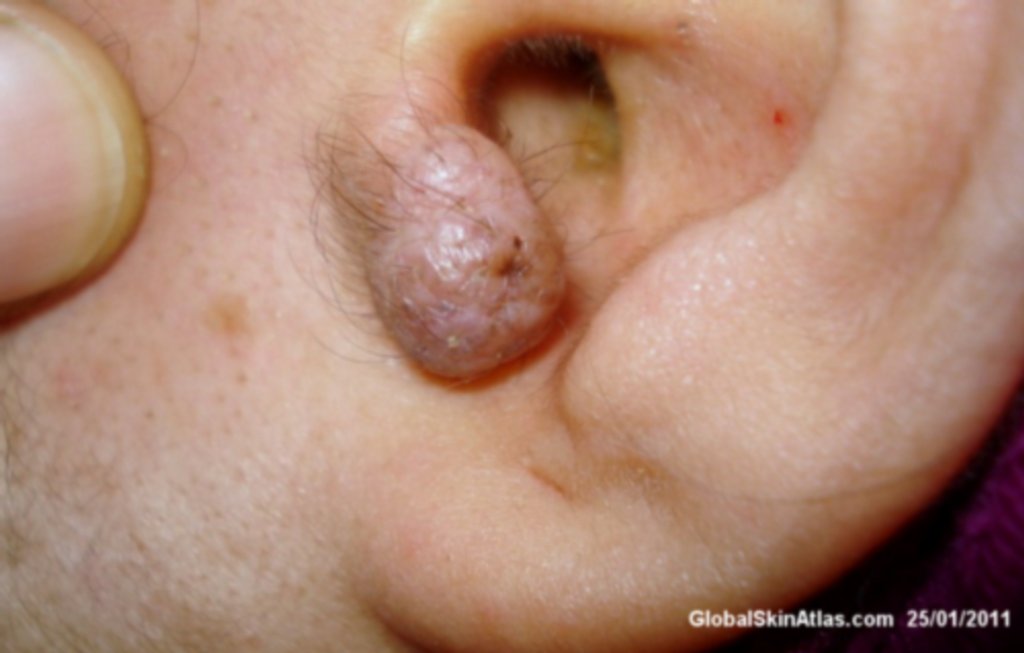
History:
Angiolymphoid Hyperplasia with Eosinophilia (ALHE) usually presents with pink to red-brown, dome-shaped, dermal papules or nodules of the head or neck, especially about the ears and on the scalp. Grouped lesions merge to form plaques or grapelike clusters. There is a female preponderance, and the average age of onset is 32 years. Symptoms can be pain or pruritus; these may occur after trauma. HistoIogically, central thick-walled vessels with hobnail endothelium are noted. Surrounding hyperplasia of smaller vessels and nodular lymphoid aggregates with eosinophils are present. Here is a good example of ALHE which has been proved histologically.