
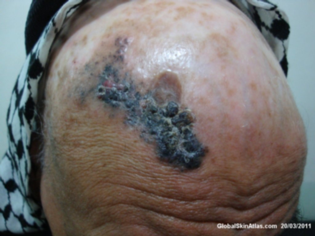
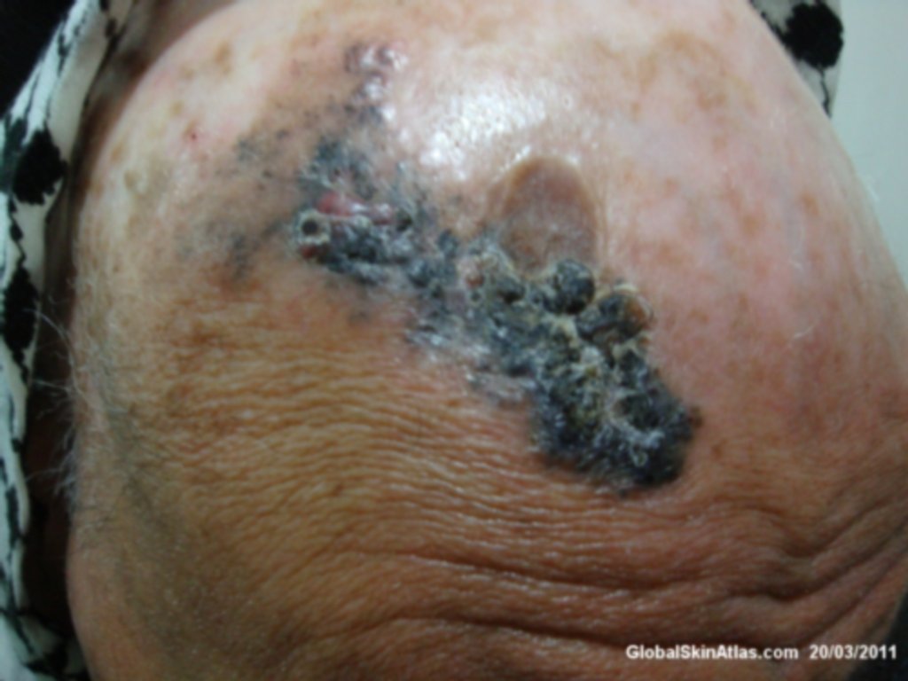
Diagnosis: Recurrent BCC
Description: Pigmented plaque with rolled up border.
Morphology: Hyperpigmentation
Site: Scalp
Sex: M
Age: 68
Type: Clinical
Submitted By: Nameer Al-Sudany
Differential Diagnosis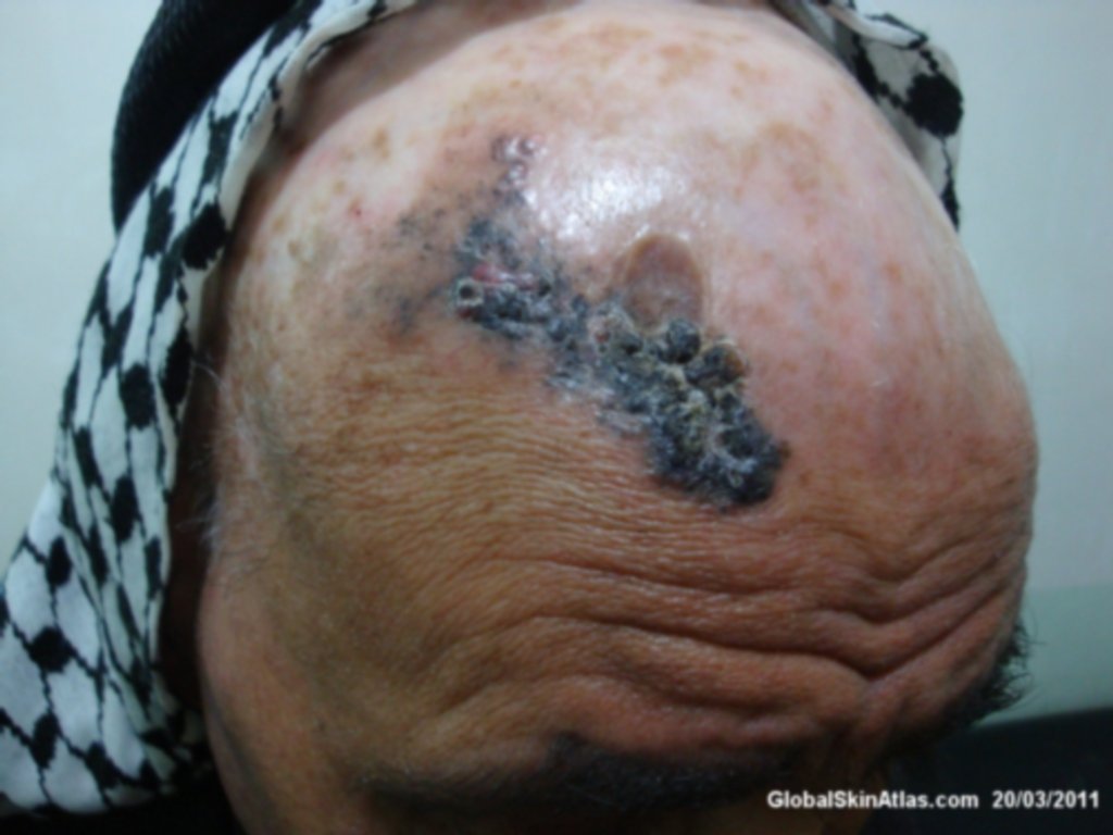
History:
At 1950s the patient was treated with radiation therapy for favus (the treatment available for tinea capitis at that time). Many decades later (1990s) he developed BCC on his atrophic scalp treated with surgical excision. Three years later it recurred and at this time it had been treated with a wrong decision from the treating dermatologist with 10 sessions radiation therapy. On radiation therapy the lesion partially disappeared. Now he presented with a large pigmented plaque having slightly raised border. Incisional biopsy showed pigmented BCC.