
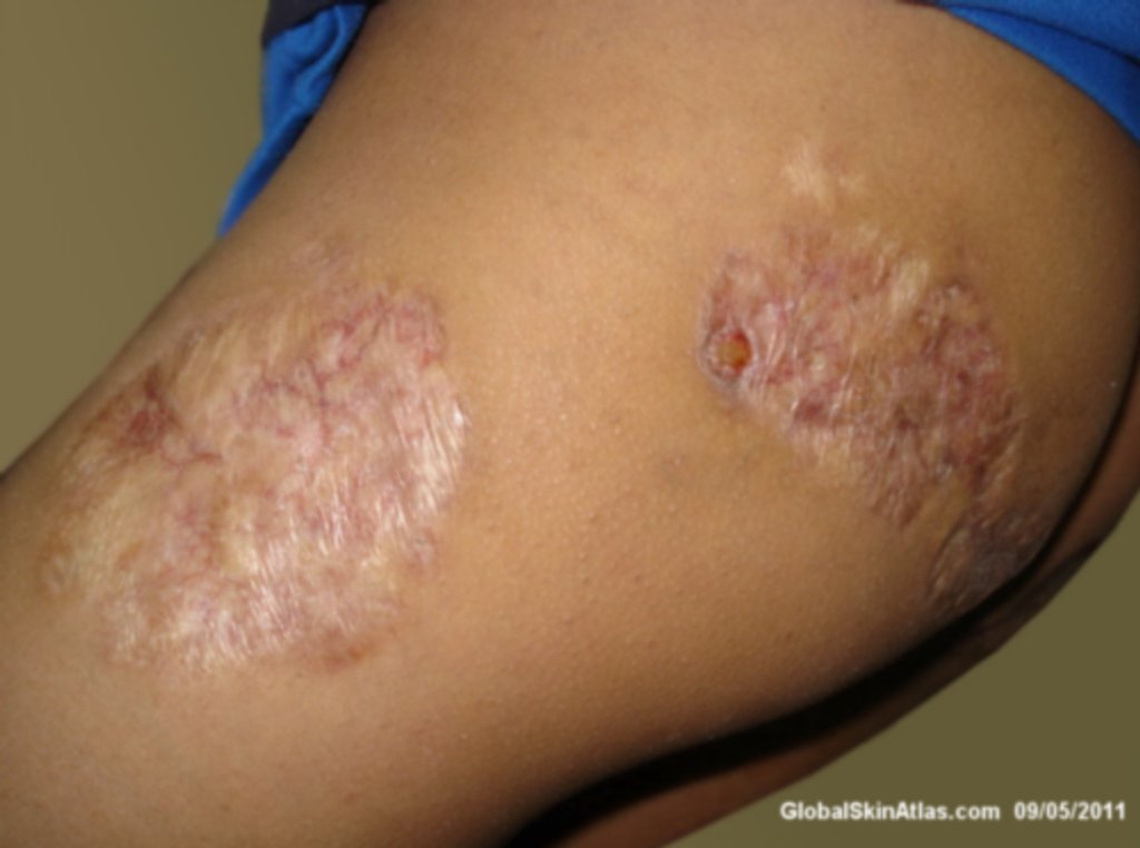
Diagnosis: Lupus vulgaris
Description: Atrophic plaques with telangiectasis.
Morphology: Atrophy
Site: Thigh
Sex: F
Age: 15
Type: Clinical
Submitted By: Nameer Al-Sudany
Differential DiagnosisHistory:
Lupus vulgaris is the most common form of cutaneous tuberculosis in many parts of the world. It occurs mostly in elderly and is usually the result of endogenous reactivation; the mycobacteria reach the dermis by direct spread from lymph nodes, or by hematogenous or lymphatic spread. It presents as large red-brown atrophic patches or plaques with telangiectases. The sites of predilection include face (especially nose and ears), breasts, and thighs. Crusts, ulceration, and destruction of adjacent tissue (cartilage of ear or nose) lead to mutilation. About 40–50% of patients have tuberculosis in other organs. Other sites include lymph nodes, mucosa, joints and bones. This girl presented with typical lesions on the thigh and buttock of more than one year duration. She had no apparent cause of tuberculosis in other areas of the body and negative family history of TB. Her skin biopsy revealed granulomas with classic three-zone pattern of tuberculosis: central necrosis, band of epithelioid macrophages and Langhans giant cells, rim of lymphocytes. No acid fast bacilli detected in skin biopsy.