
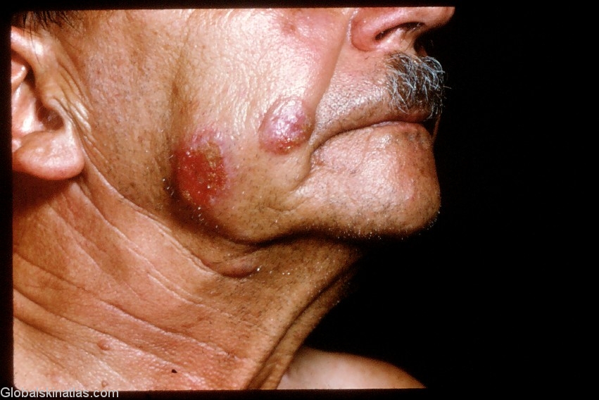
Diagnosis: Follicular Mucinosis
Description: Large plaques of mucin deposition in the skin
Morphology: Plaque
Site: Cheek
Sex: M
Age: 68
Type: Clinical
Submitted By: Ian McColl
Differential Diagnosis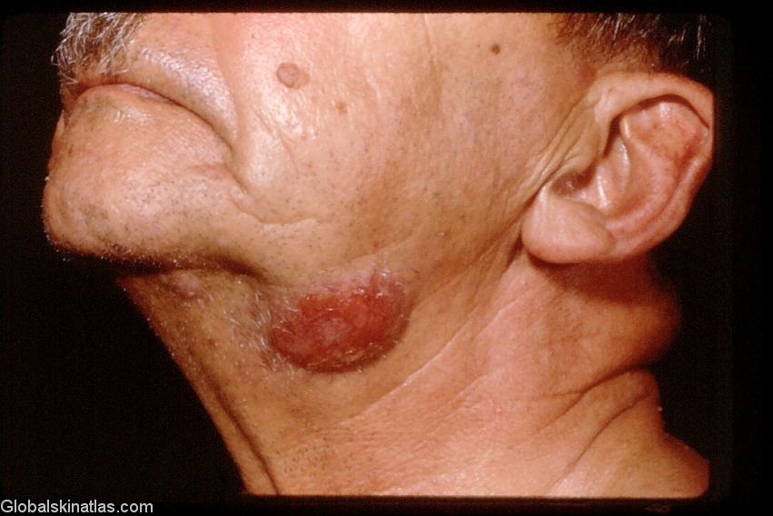
History:
This man was one of the rarer cases of Follicular mucinosis which are associated with Mycosis fungoides.Pressure on the plaques would cause clear mucin to bead up from the hair follicle openings although this phenomenon is commoner in the benign types.His histology showed epidermotropism and atypical lymphocytes.
The folowing reference in Dermatology Journal Online describes a case with these characteristics.