
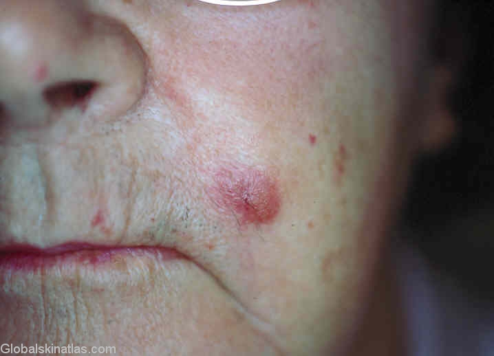
Diagnosis: Lymphocytoma cutis
Description: Red slightly scaly nodule on the cheek
Morphology: Nodule pink
Site: Cheek
Sex: F
Age: 62
Type: Clinical
Submitted By: Ian McColl
Differential DiagnosisHistory: Lymphocytoma cutis is usually described as a red or purplish nodule or plaque in the skin secondary to an insect or tick bite.The earlobes are a characteristic site particularly in children.In some parts of the world it is due to Borrelia infection and may be a manifestation of Lyme disease.However they bear watching because early lymphoma cutis may present in a similar fashion .T cell gene rearangement studies looking for monoclonality will separate out this variant.This lady's lesion responded to some intralesional steroid.She had no evidence of Borrelia infection.