
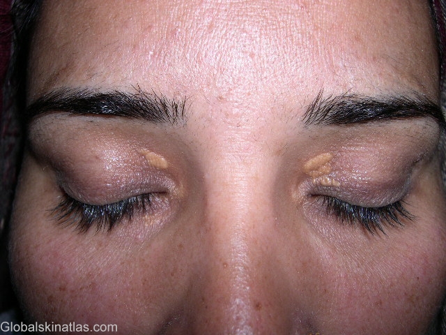
Diagnosis: Xanthelasma
Description: Yellow plaques
Morphology: Plaque
Site: Eyelids
Sex: F
Age: 45
Type: Clinical
Submitted By: Shahbaz Janjua
Differential Diagnosis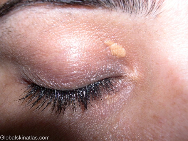
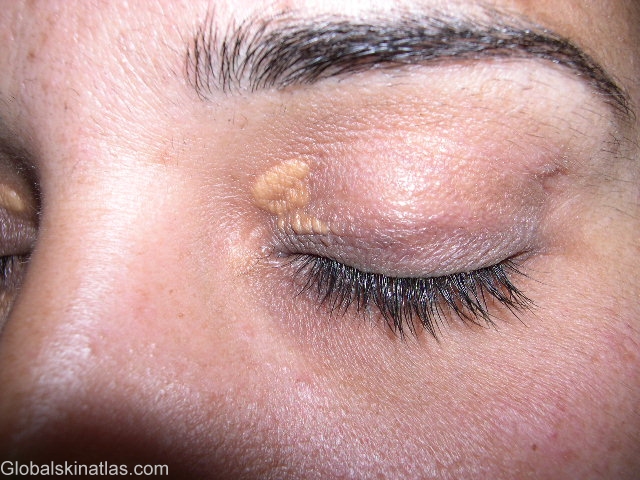
History: Xanthelasma or xanthoma palpebrarum are yellow plaques that are usually located on the medial side of the upper eyelids. One half of these lesions have been found to be associated with elevated plasma lipid levels. Histopathological examination shows xanthelasma to be composed of xanthoma cells which are foamy histiocytes laden with intracellular fat deposits of cholesterol primarily within the upper reticular dermis.