
Diagnosis: Onychomycosis
Description: Overview
Morphology: Nail dystrophy
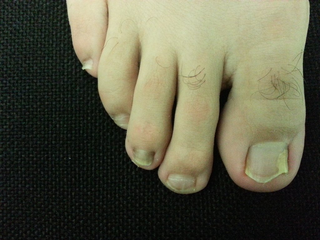
Site: Toes
Sex: M
Age: 27
Type: Clinical
Submitted By: Goh Robin Yeong Hong
Differential Diagnosis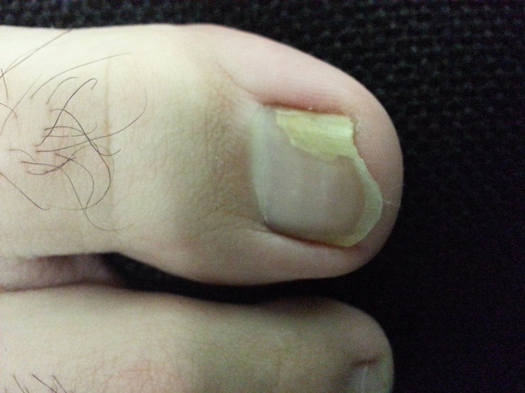
History:
27 healthy male otherwise healthy
Complains of new development of nail dystrophy associated with slight pain.
after several months of soccer and trauma to foot.
GP given lamasil topical but failed therapy .
o/e.
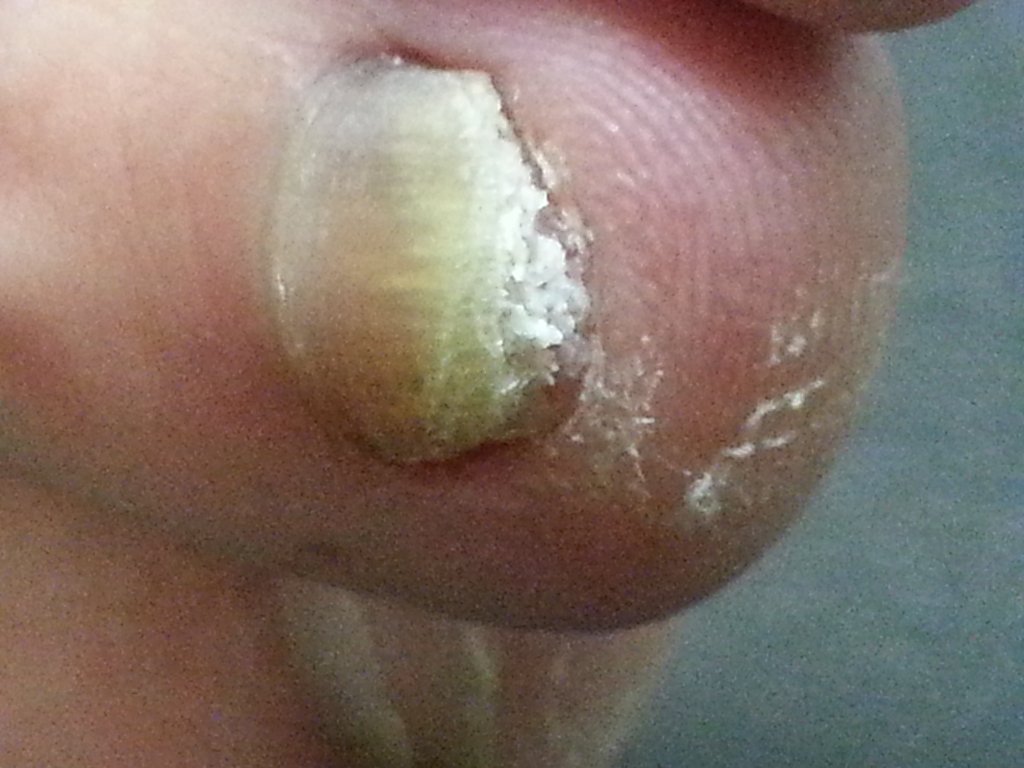
1st toe: Distal lateral subungal yellowing
3rd toe: Hyperkeratosis of nail bed.
Absence of paronychia.
Investigations:
Fungal examination (KOH examination) & PAS Histopathology
1. Nail clippings microscopy of 1st digit
Non specific fungal elements seen. Distinction between yeast, dermatopyte or other fungus cannot be made.
Culture: Trichophyton rubrum
2. Nail Clippings microscopy of 3rd digit
Fungal elements NOT seen.
Culture: Trichophyton rubrum
2. PAS Histopathology of 1st and 3rd digit.
- Histopathology: Fungal bodies were positive in large numbers.
Management:
Have advised patient it might not be necessary to treat.
Patient is keen to try oral therapy.
Prior to treatment: FBC, LFT were all within normal range.
Patient given oral terbinafine 250mg OD for 3 months.
Comments:
T. rubrum is the most common causative pathogen for onychomycosis.