
Diagnosis: Leishmaniasis
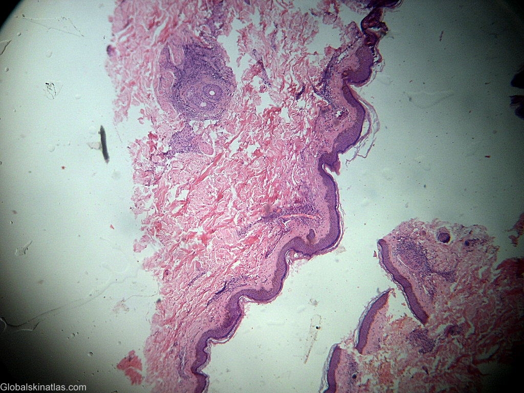
Description: Dense mixed inflammatory cell dermal infiltrate.
Morphology: Path,superficial pvd
Site: Face
Sex: M
Age: 35
Type: Clinical & Histology
Submitted By: Shahbaz Janjua
Differential Diagnosis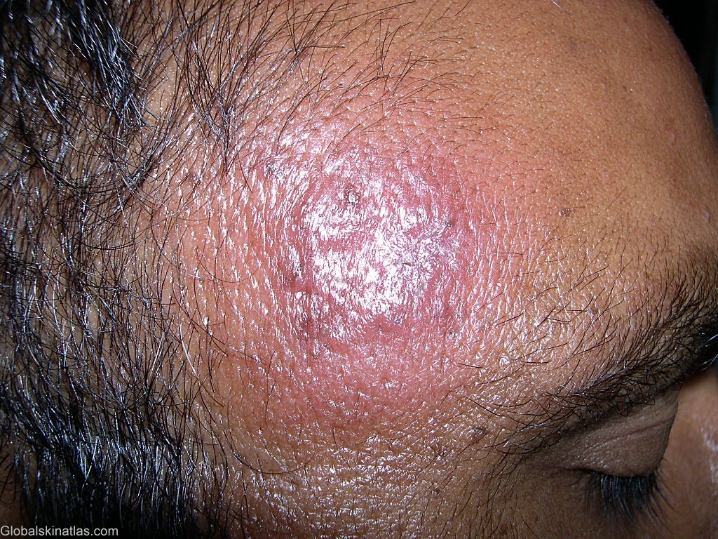
History:
A dense mixed inflammatory cell infiltrate in the superficial dermis comprising of lymphocytes, histiocyes, neutrophils, plasma cells and epithelioid cells is not seen in other chronic granulomatous diseases. Hence, it should be considered diagnostic of cutaneous leishmaniasis even in the absence of LT bodies.
Reference: Mashhood AA et al. Fine needle aspiration cytology versus histopathology in the diagnosis of cutaneous leishmaniasis in Pakistan. J Coll Physicians Surg Pak. 2000 Feb;15(2):71-3.