
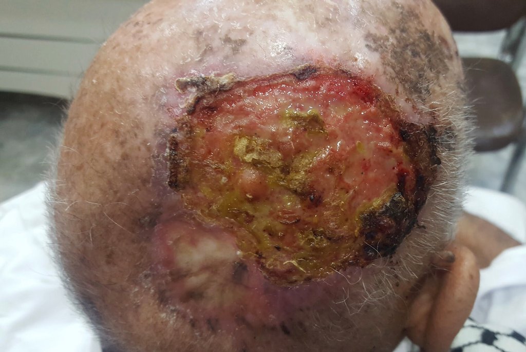
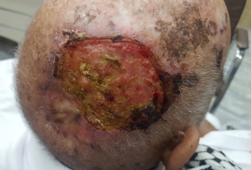
Diagnosis: Basisquamous carcinoma
Description: Giant ulcer
Morphology: Ulcer
Site: Scalp
Sex: M
Age: 82
Type: Clinical
Submitted By: Nameer Al-Sudany
Differential Diagnosis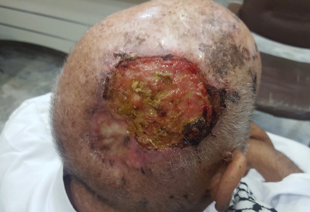
History:
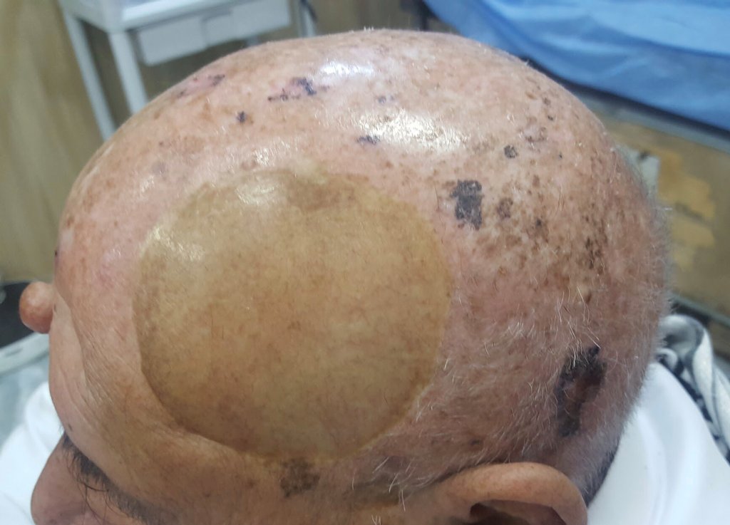
This elderly man had exposed to radiation therapy for tinea capitis in 1950s; he presented with a large ulcer having an everted border of about one year duration. The new ulcer arouse from a site of previously treated non melanocytic epidermal tumor (BCC or SCC ?). O/E, many pigmented BCCs, actinic keratoses and a prvious large BCC, which was treated sugically and skin grafted two years ago (see last photo), were seen on an atrophic scalp! Incisional biopsy wa taken from the border of the new recurrent giant ulcer which showed a BASISQUAMOUS CA on histological examination.