
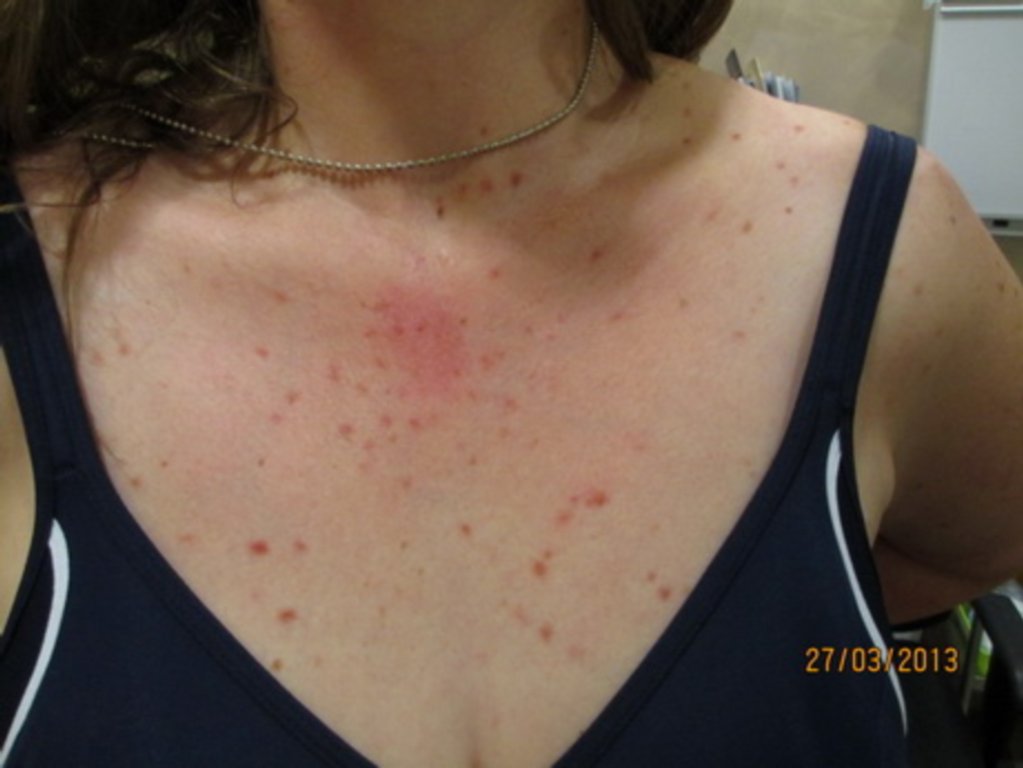
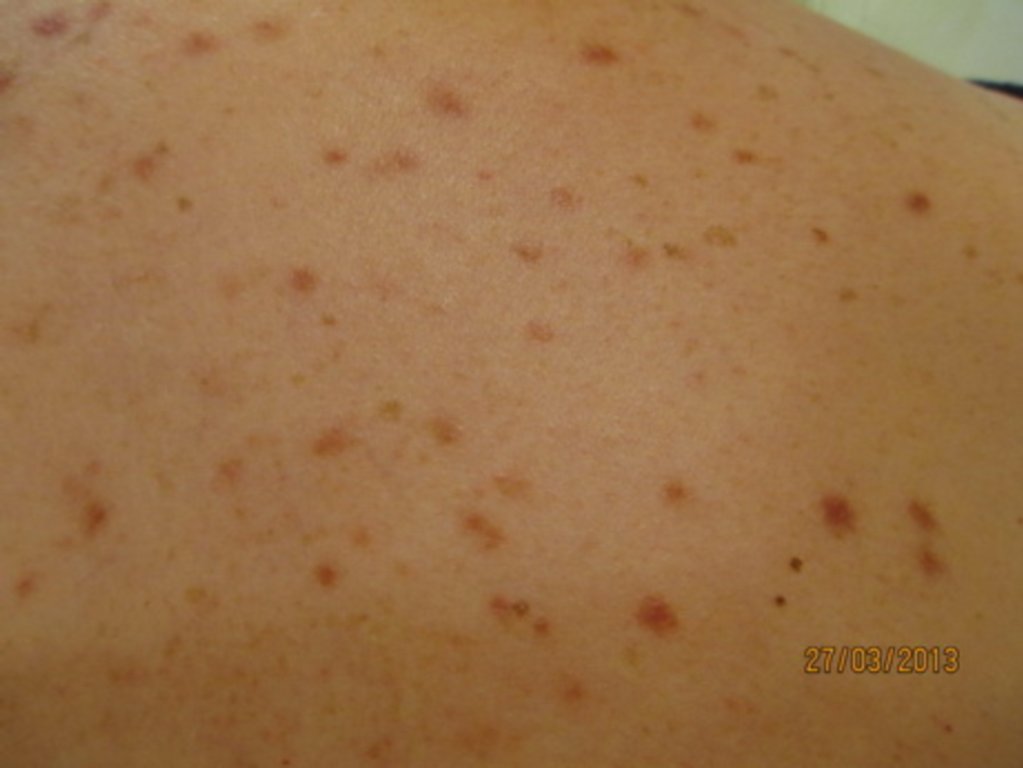
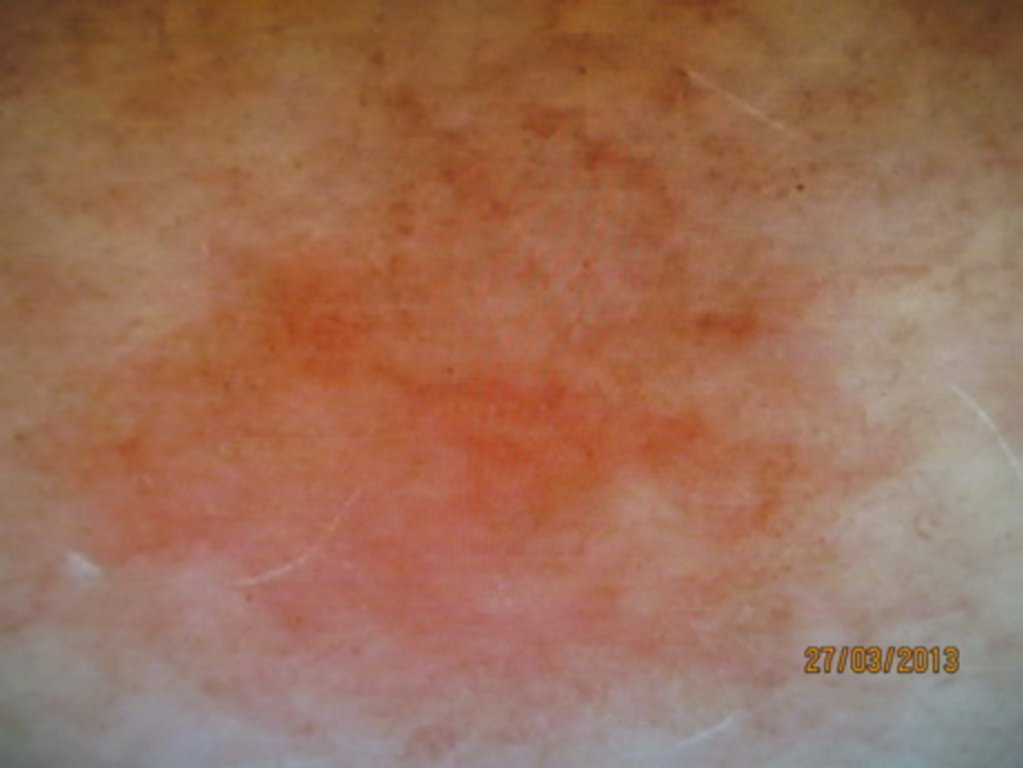
Diagnosis: Urticaria Pigmentosa
Description: Brown macules and red urtication from histamine release when rubbed
Morphology: Macule brown
Site: Chest
Sex: F
Age: 40
Type: Clinical
Submitted By: Ian McColl
Differential Diagnosis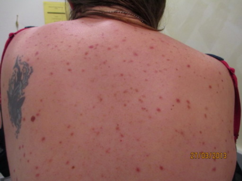
History:
40 year old women presented with 3-4 yerars history of pruritic rash onm trunk and limbs, Face, scalp, mucosa, palms and soles spared. Rash follows the same pattern: pruritic red macule that she scratches, which evolves into red nodul and after several weeks that changes into hyperpigmented flat papule or macule, which persist.
Generally she has been well, no past medical histroy, no significan family history, not on medication. Uses sun protection.
Blood tests including autoimmune screen were normal.
Biopsy revealed following findings:
Section of the skin shows mild hyperplasia of the epidermis, basal hyperpigmentation and mild compact orthokeratosis. This is associated with light perivascular lyphohystiocytic infiltrate with occasional eosinophils, and an infiltrate of mast cells in the superficial dermis.
The Mast cells stain for CD117 and chloroacetate esterase. No fungal infection on PAS stains or malignancy is identified.
Direct immunofluorescence studies for IgG, IgM IgA and C3 are negative.
Comment: The appearances are those of mastocytosis and are in keeping with urticaria pigmentosa. Since patient is an adult, systemic disease should be excluded.