Loading
Gloal Skin Path Atlas-logo
  • Recent Submissions
  • Search
  • Diagnosis
  • Quiz
  • Submit
  • Index
  • Blog
  • About GSA

<< Back to Diagnosis Page

Image Number #8719 (Merkel cell carcinoma)

Diagnosis: Merkel cell carcinoma

Description:

Nodule selected for excision biopsy

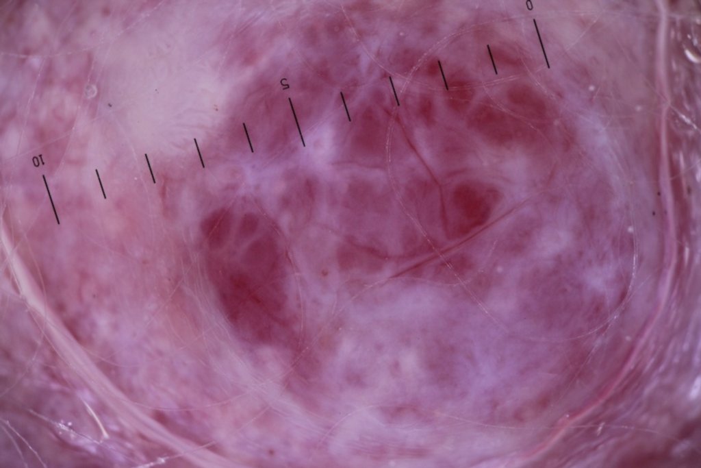
Morphology: Nodule pink
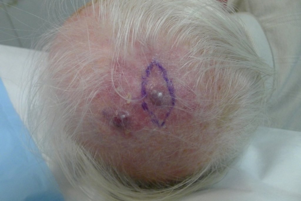
Site: Scalp

Sex: M

Age: 81

Type: Clinical & Histology

Submitted By: Ian McColl

View Full Size Image

Differential Diagnosis
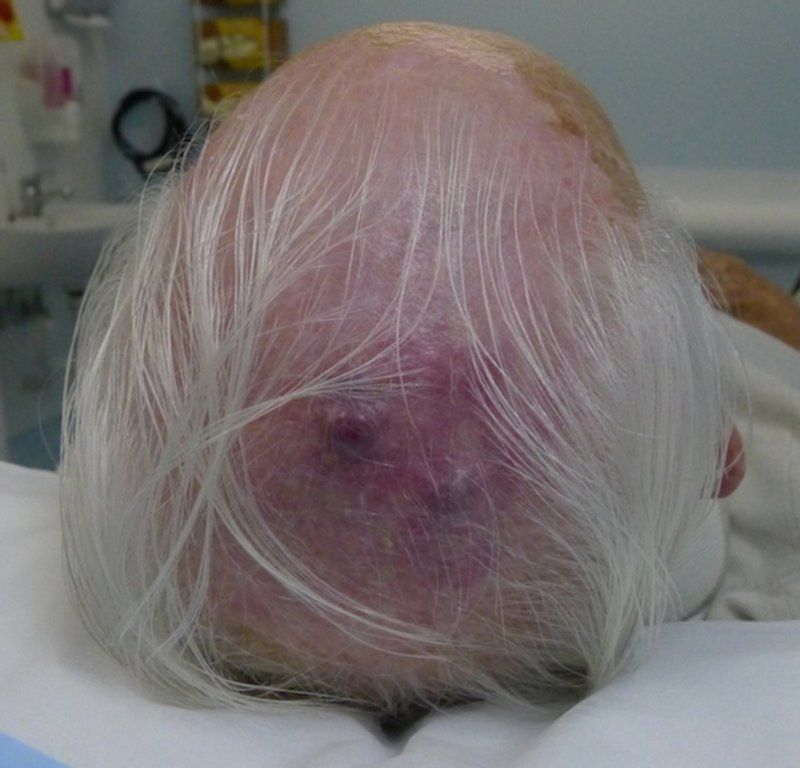
Click Here to View Details

History:

Case from Dr 

81yo male (last seen 6/12 ago) presents for routine skin cancer examination and has noted a 2/12 history of growing nodules on his scalp. He visited his regular GP a few weeks earlier and mentioned them. His GP allegedly said that these lesions were benign and would need laser removal.   Excision would bleed too much so, not worth doing.

PMhx: Well. Minimal medication. Increased PSA, monitored by Urologist only at this stage.
Has had surgical treatment of superficial SCC and BCC's in past

Full systems review revealed no concerning symptoms with regards to sinister internal malignancies

OE:
3 relatively large pink nodules surrounded by pink erythematous telangiectatic area of approximately 10cm diameter.

Dermoscopically: featureless pink clods with white lines and white featureless areas. They were much more featureless than I had expected macroscopically. Haemangiomas in general have pink/red or purple clods mainly with limited white lines.

General examination normal with head, neck or supraclavicular lymphadenopathy

Imp: Fast growing pink nodules on a solar damaged scalp of an elderly gentleman.

Dx: Merkels Cell Carcinoma
Ddx: Cutaneous metastasis (?SCC), amelanotic melanoma, atypical fibroxantoma, pyogenic granulomas, haemangioma.

I excision biopsied an entire nodule from the field for diagnosis. The diagnosis was 'strongly suggestive of Merkel Cell Carcinoma' and involved the entire specimen thickness and the margins.

Mx: Urgent referral to radiation oncologist last week, booked plastic surgeon for wide margin excision with skin graft. PET scan today revealed no spread to organs at this stage. Will follow up in 3/12 after surgery and radiation treatment.

Follow Up
histo report for those with an interest:

Clinical notes:   Central scalp - Rapid growing large pink nodules x 3 in
the field, not present 3/12 ago. ?metastasis (?SCC), merkel cell ca,
amelanotic MM, atypical fibroxanthoma. Excision of just one nodule at this
stage for diagnosis, Photos taken if required.

Macroscopic:

The specimen consists of an ellipse of skin measuring 29 x 16 x 3mm bearing
a pale pink papule measuring 10mm in diameter. Towards one apex of the
specimen there is an orientation nick present. As no orientation is
specified, this is arbitrarily placed towards the 12 o'clock margin. The 3
o'clock margin is scored. Starting from the 12 o'clock margin moving
towards 6 o'clock, 3TS block A and 3TS block B. pp/ln

Microscopic:

Sections show an undifferentiated small round cell malignancy which is
present in severely sun-damaged skin. The lesion comprises multiple often
coalescent nodules and diffuse areas. The tumour involves the superficial
dermis to deep fascia. The cells have minimal cytoplasm and round to oval
hyperchromatic nuclei with indistinct nucleoli and coarse granular
chromatin. There is frequent mitotic activity. An epidermal component is
not identified. Focal lymphatic permeation is seen. There is no
perineural invasion. The lesion broadly involves the deep and peripheral
margins of this specimen. Immunohistochemistry will be performed to
further characterise the nature of the malignancy.

SUMMARY:
SCALP - UNDIFFERENTIATED SMALL ROUND CELL MALIGNANCY. LESION INVOLVES THE
PERIPHERAL AND DEEP MARGINS. IMMUNOHISTOCHEMISTRY IS PENDING.


FURTHER REPORT ADDED 20 APRIL 2015

Immunohistochemistry has been performed. The tumours cells are strongly and
diffusely positive for AE1/AE3, Cam 5.2, Synpatphysin and CD56. They are
weakly positive for Chromogranin and negative for leukocyte common antigen,
S-100 protein, HMB45 and thyroid transcription factor 1. CK7 and CK20 are
both strongly positive in most cells with a paranuclear dot present in a
many of the cells.

The immunohistochemical features are not completely specific but are most
in keeping with MERKEL CELL CARCINOMA. A metastatic small cell
neuroendocrine is still possible but considered less likely. The negative
result for TTF-1, while not excluding a metastasis from lung primary, makes
it less likely.

SUMMARY:
SCALP - FEATURES IN KEEPING WITH MERKEL CELL CARCINOMA. PERIPHERAL AND DEEP
MARGINS INVOLVED.


DermNetNZ   eMedicine   PubMed   Dermatology Online   Archives   JAAD for "Merkel cell carcinoma"

  • Recent Submissions
  • Search
  • Diagnosis
  • Quiz
  • Submit an Image
  • Index
  • About GSPA

Copyright 2022. Global Skin Atlas. ALL Rights Reserved.

Change Image