Loading
Gloal Skin Path Atlas-logo
  • Recent Submissions
  • Search
  • Diagnosis
  • Quiz
  • Submit
  • Index
  • Blog
  • About GSA

<< Back to Diagnosis Page

Image Number #8801 (Dermatophytosis)

Diagnosis: Dermatophytosis

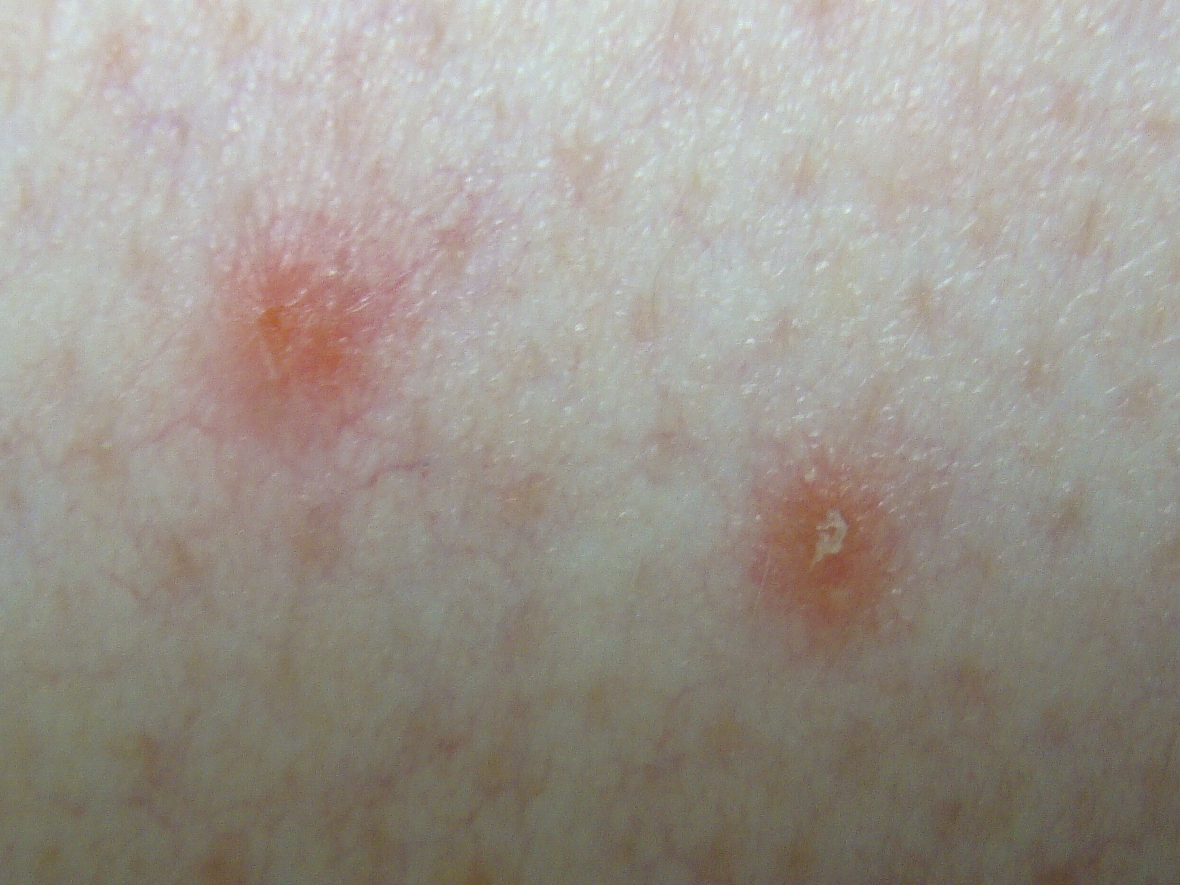
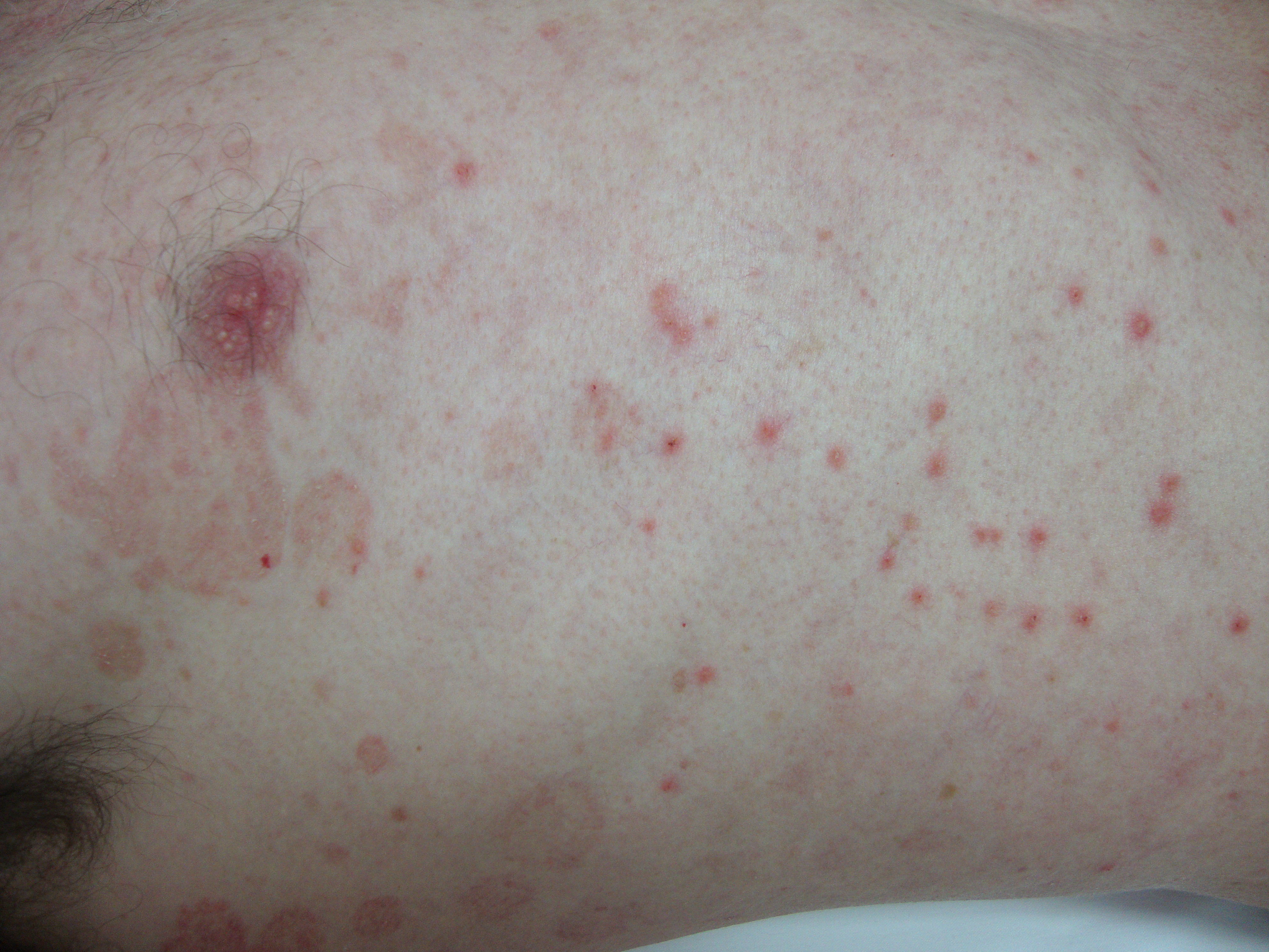
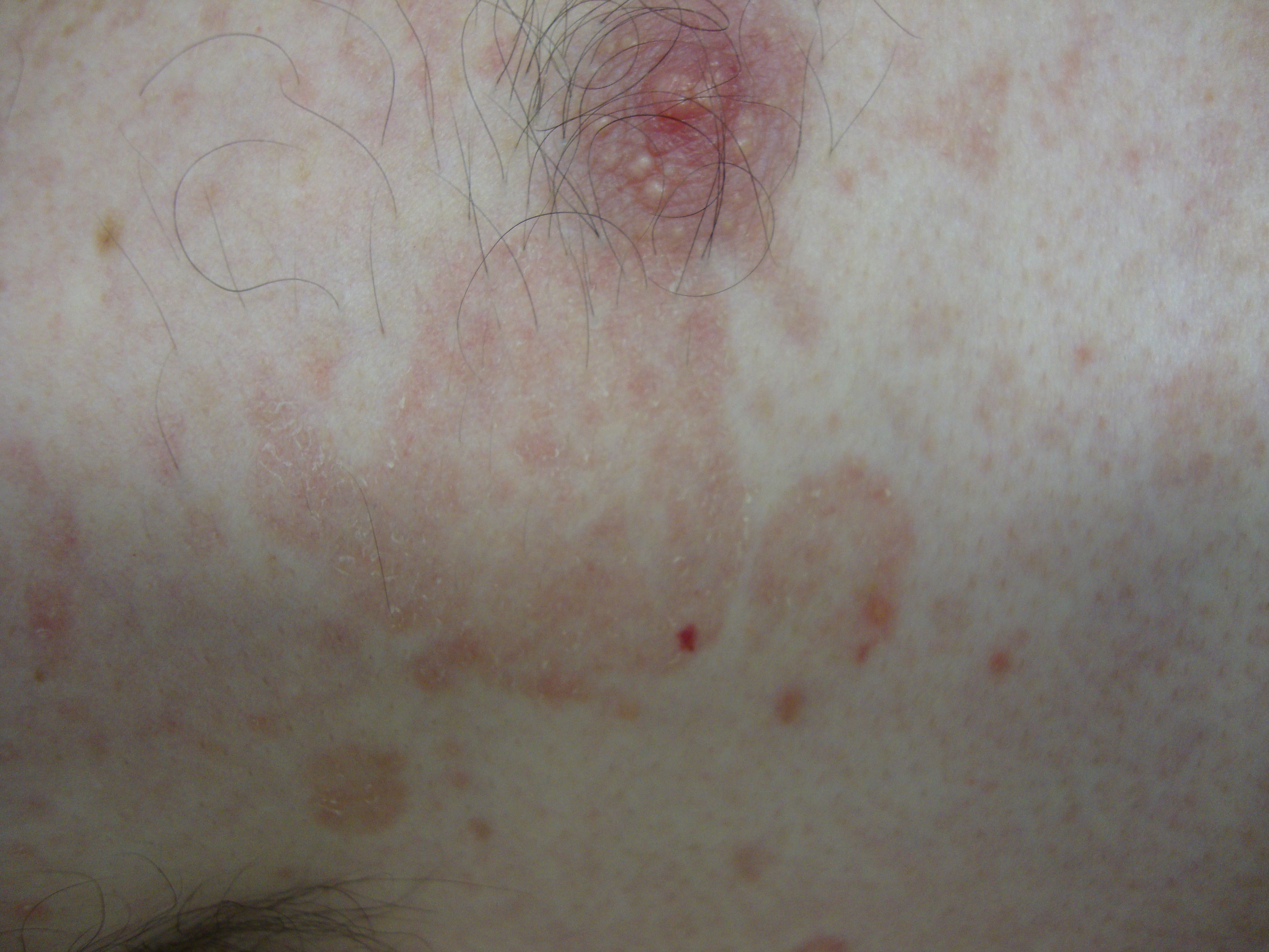
Description:

Inflammed scaly papule rather than pustule

Morphology: Red,scaly

Site: Chest

Sex: M

Age: 34

Type: Clinical

Submitted By: Ian McColl

View Full Size Image

Differential Diagnosis
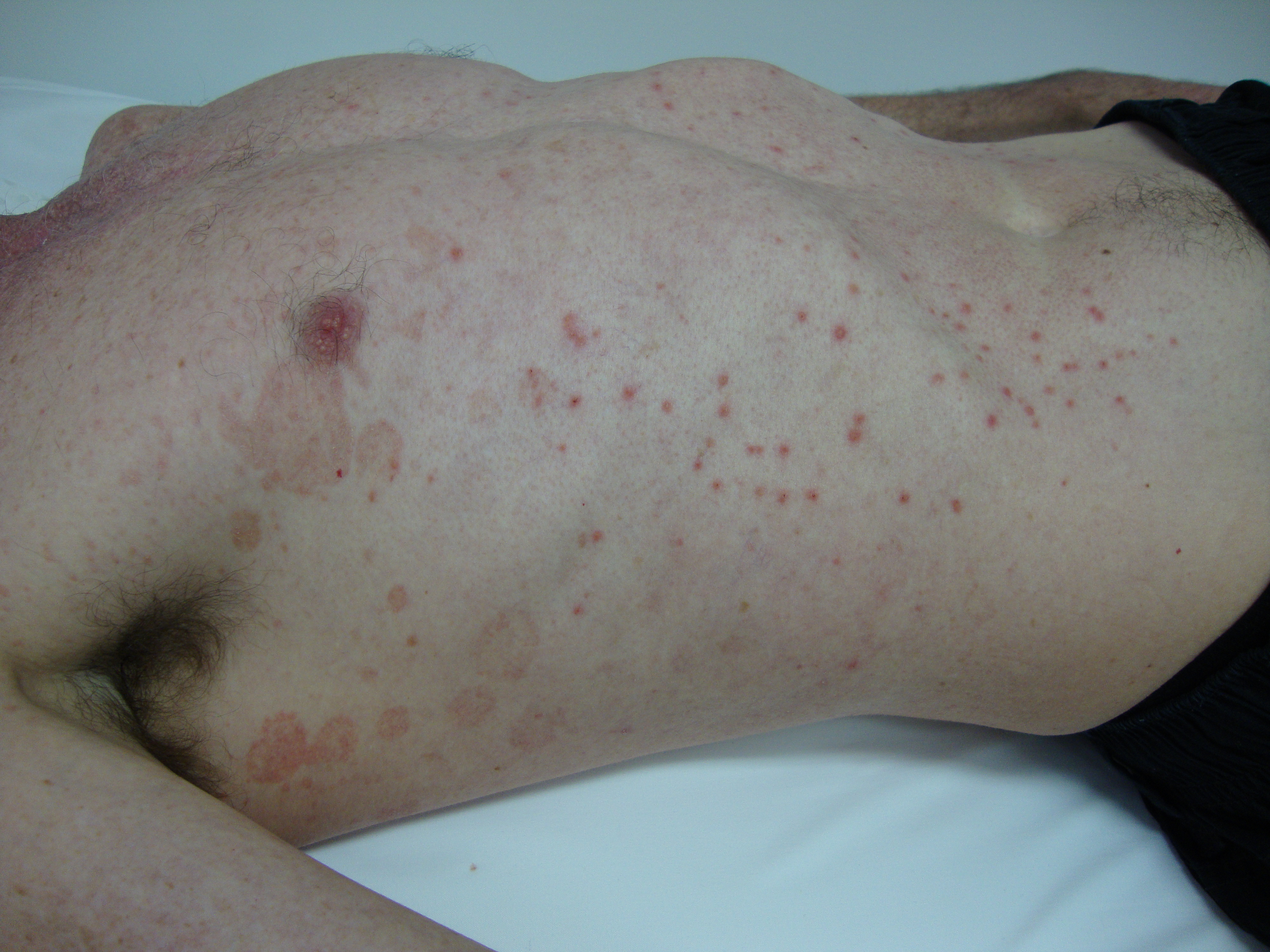
Click Here to View Details

History:

Patient of Dr 

34 yo  , no sig PMHx / FHx , no med . 12/12 of pruritic erythematous dry rash. Clearly he seemed to me to have tinea but also he had another rash which I was not clear what it was Was I dealing with two pathologies ? were they related . Bx showed Tinea and acantholytic keratosis as being the pustular component of the rash . What is the relation and how to manage

Clinical Notes: Biopsy Report :

12 Months of itchy red scaly rash.
1. Large erythematous patch / looks tinea.
2. Pustular lesion.

Macroscopic:
1. 'Axilla'. The specimen consists of two 2mm punch biopsies.

2. 'Trunk'. The specimen consists of a punch biopsy of skin
measuring 3mm in diameter and 3mm in depth. (DM/DT)

Microscopic:
1. The biopsy shows skin with epidermis and dermis. The PAS stain
shows fungal elements on the surface. In the dermis there is a patchy
perivascular lymphocytic infiltrate. The features are in keeping with
the history of tinea.

2. The biopsy shows an acantholytic lichenoid keratosis. Multiple
levels have been examined. No microscopic pathogens are seen.

Summary:

1. AXILLA - IN KEEPING WITH DERMATOPHYTE / TINEA.
Specimen No : 10869-09SP
2. TRUNK - ACANTHOLYTIC KERATOSIS.

When I looked at the distant view I thought he had tinea versicolor plus an excoriated dermatitis but the scale on the close up is more a dermatophyte than a yeast. Did you scape it for culture and microscopy? Remember a skin scraping is a better way to confirm a fungal infection than a skin biopsy!
Also the acantholytic histology of the other biopsy suggests he also has Grover's syndrome, a condition like Tinea versicolor which is also associated with excess sweating. It will respond to a topical steroid such as Celestone M cream (Betamethasone 0.02%) but of course this would not be the best thing for either Tinea corporis or Tinea versicolor! If Tinea versicolor the oral Nizoral is best and either Griseofulvin or Terbinafine orally for tinea corporis. That is why the scrapings and culture are important! (Submitted By: Dr Ian McColl)


DermNetNZ   eMedicine   PubMed   Dermatology Online   Archives   JAAD for "Dermatophytosis"

  • Recent Submissions
  • Search
  • Diagnosis
  • Quiz
  • Submit an Image
  • Index
  • About GSPA

Copyright 2022. Global Skin Atlas. ALL Rights Reserved.

Change Image