
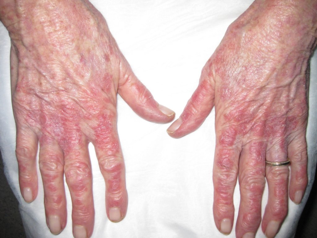
Diagnosis: Subacute lupus erythematosus
Description: Note the distribution of the erythema between the joints
Morphology: Red,nonscaly
Site: Hand,dorsum
Sex: F
Age: 82
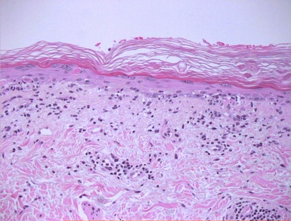
Type: Clinical & Histology
Submitted By: Ian McColl
Differential Diagnosis
History:
Patient of Dr
82 yo female presented with this rash on her face, neck and forearms in light-exposed areas 2 weeks after receiving an intravitreal injection for treatment of her macular degeneration. The rash began approx 5 days post-injection as a few "flat, brown spots" and then evolved into the rash seen below. Biopsy showed a prominent lichenoid inflammatory process with some necrotic keratinocytes in keeping with a drug reaction and, considering the clinical history, consistent with the diagnosis of drug-induced, subacute cutaneous lupus erythematosus.
This is the first documented evidence of an intravitreal injection of a monoclonal antibody causing a systemic reaction like SCLE.
We plan to publish this as a case report.
usually we see this as a reaction to lucentis for ARMD (age related mac degeneration) if the patient goes in the sun (Submitted By: Dr Leroy Rebello)