Loading
Gloal Skin Path Atlas-logo
  • Recent Submissions
  • Search
  • Diagnosis
  • Quiz
  • Submit
  • Index
  • Blog
  • About GSA

<< Back to Diagnosis Page

Image Number #8992 (Disseminated superficial actinic porokeratosis)

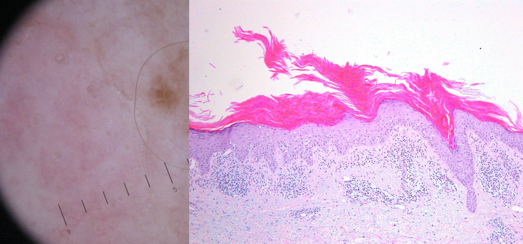
Diagnosis: Disseminated superficial actinic porokeratosis

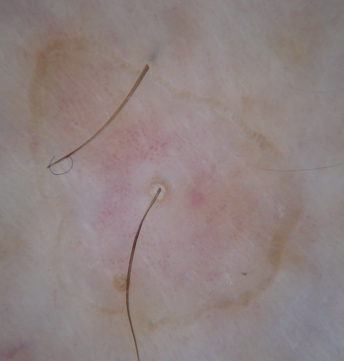
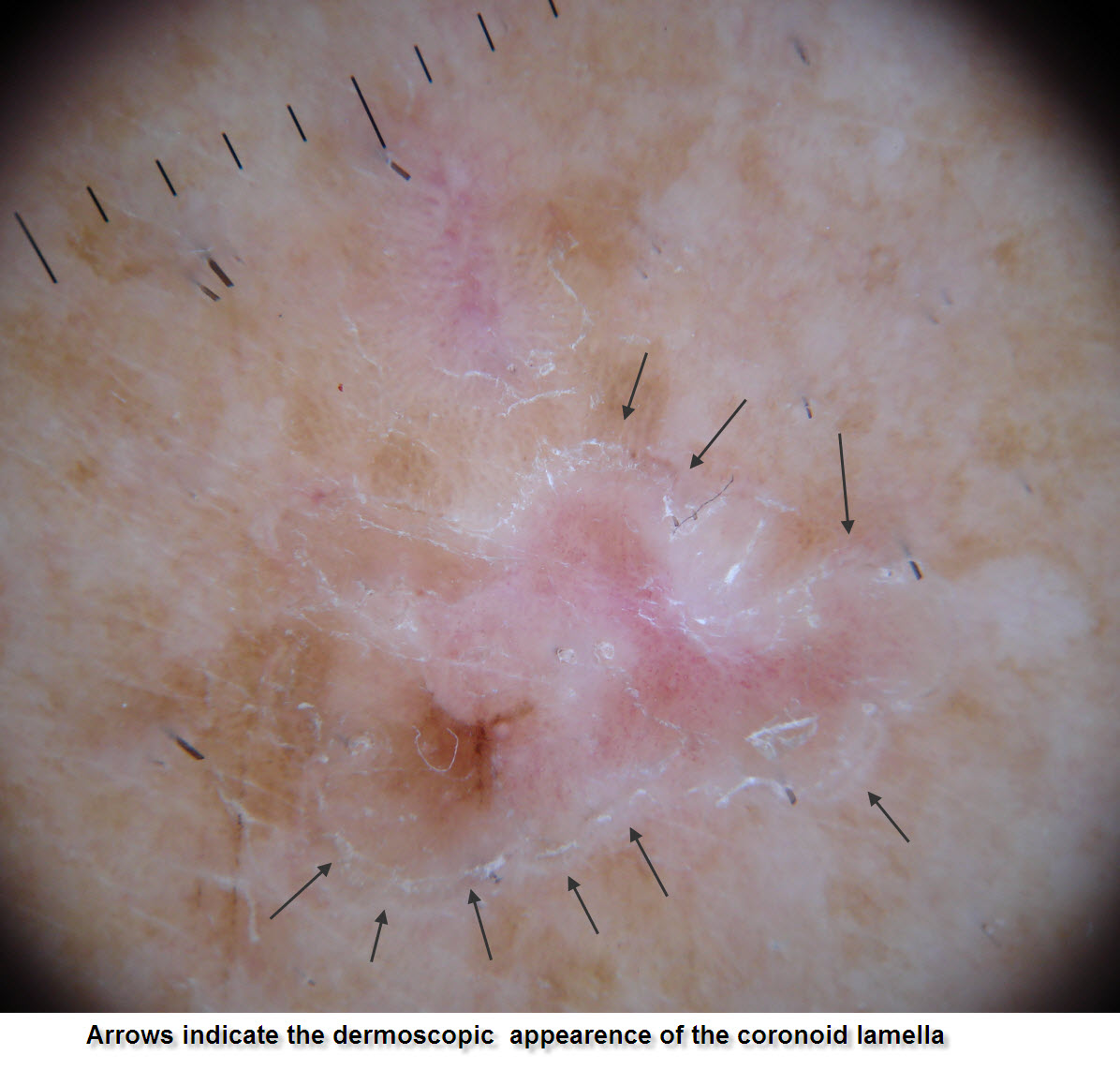
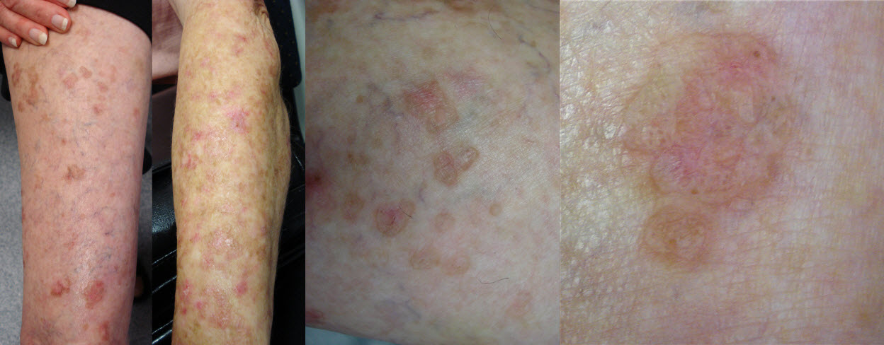
Description:

Porokeratosis of Mibelli

Morphology: Red,scaly

Site: Limbs

Sex: F

Age: 70

Type: Clinical & Histology

Submitted By: Ian McColl

View Full Size Image

Differential Diagnosis
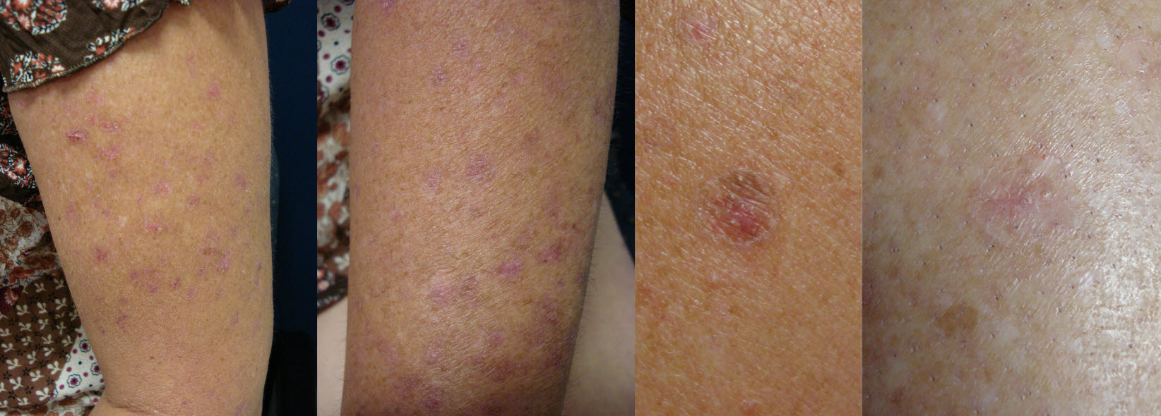
Click Here to View Details

History:

Case 2_G.C.

There are actually 3 patient’s presented here a mother and daughter (aged 70's and 40's) with multiple erythrosquamous lesions on the arms and legs, and a third patient (female late 50’s)with a solitary large annular erythrosquamous plaque on the distal triceps. These cases illistrae one the small "s" (solar damage) conditions in the PMsPET pneumonic.

The mother and daughter have typical disseminated superficial actinic porokeratosis (DSAP) one of a group of Porokeraoses. The final case is Porokeratosis of Mibelli. .

In both clinically the typical finding is an annular rim of keratin scale which histollogically is described as a coronoid lamella.

Treatment can be problematic the 2 girls with DSAP have had multiple lesions treated with cryotherapy or Efudix, but other new lesion appear to replace those treated. UV protection is paramount and there is as with solar keratoses a small risk of lesions progressing to IEC/SCC. 80% Liquid phenol apparently works well but I have yet to try this. The isolated Mibelli lesion is being treated with efudix , I will post some follow up images in due course.


Related Links:
Disseminated superficial actinic porokeratosis
Porokeratosis of Mibelli
 

Case Comments     [Add Comment] [Subscribe]
This is an excellent example of this condition. Disseminated superficial actinic keratoses present as a red scaly rash on the arms and legs usually having been there for some time but flaring after sun exposure. Once you spot the castle rampart line surrounding the lesion you have made the diagnosis. A common mistake is to biopsy the red central part of a lesion and get a lichenoid histological pattern and have it reported as lichen planus or lichenoid keratosis depending on the information you have given the pathologist. You have to biopsy the surrounding rim to show the cornoid lamella which is the hallmark of these lesions. Inherited as autosomal dominant ie half the children inherit it but it takes 20 odd years of sun exposure before the lesions start to come out and curiously they spare the face!   
(Submitted By: Dr Ian McColl)

    [Modify]

Treatment This is difficult. 80% phenol sounds like it will leave multiple hypopigmented spots if you use it. Sun protection is most important and by doing just this the redness can settle down although the cor noid lamellae remain but the rash is less obvious. I freeze early lesions and use efudix cream bd for 4 weeks on others, tried PDT and Aldara but it is a slow process. Generally once they understand what it is and take adequate sun protection the cosmetic aspect is not so bad. The risk of scc developing is very small but scc in situ can occur. Watch one that is more inflammed or thicker than the others and biopsy it centrally, Either surgically excise or treat with Imiquimod (Aldara) for 6 weeks. (Submitted By: Dr Ian McColl)

DermNetNZ   eMedicine   PubMed   Dermatology Online   Archives   JAAD for "Disseminated superficial actinic porokeratosis"

  • Recent Submissions
  • Search
  • Diagnosis
  • Quiz
  • Submit an Image
  • Index
  • About GSPA

Copyright 2022. Global Skin Atlas. ALL Rights Reserved.

Change Image