
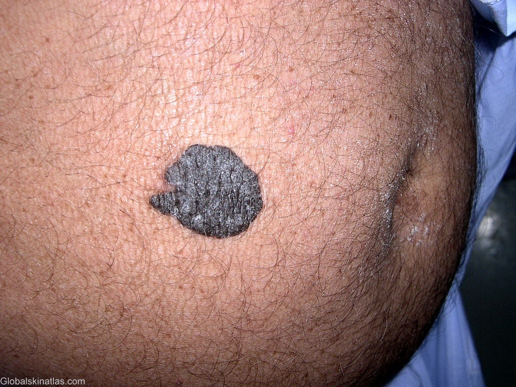
Diagnosis: Seborrhoeic keratosis
Description: A sharply defined solitary lesion
Morphology: Warty
Site: Abdomen
Sex: M
Age: 60
Type: Clinical
Submitted By: Shahbaz Janjua
Differential DiagnosisHistory:
Seborrheic keratoses are the most common benign tumors in older individuals. They develop from the proliferation of epidermal cells and their etiology remains unknown. They can occur on almost any site of the body, with the exception of the palms and soles and mucous membranes. Dermatosis papulosa nigra, stucco keratosis, and melanoacanthoma are variants of seborrheic keratosis. Differential diagnosis of seborrheic keratoses includes malignant melanoma, melanocytic nevus, verruca vulgaris, condyloma acuminatum, fibroepithelial polyp, epidermal nevus, actinic keratoses, pigmented basal cell carcinomas, and squamous cell carcinomas.