
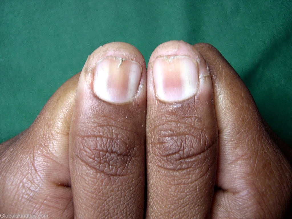
Diagnosis: Longitudinal erythronychia
Description: Longitudnal erythematous band
Morphology: Nail dystrophy
Site: Nails
Sex: F
Age: 25
Type: Clinical
Submitted By: Shahbaz Janjua
Differential DiagnosisHistory:
Longitudinal erythronychia is a longitudinal streak or band in the nail plate commencing from within the matrix and running to the point of separation of the nail bed and nail plate. At this location, the distal margin of longitudinal erythronychia may be marked by a small keratosis, arising from the nail bed and adherent to the undersurface of the nail plate. Proximal to this, there may be splinter haemorrhages in the nail bed. Beyond this point the nail plate may split in line with the erythronychia, presenting a "v"-shaped nick in the free edge. When nails are long, this sign is more prominent.
Longitudinal erythronychia may present as a single or paired band in a single nail or as multiple bands in several nails. As a single band it is likely to reflect a focal matrix pathology, such as a benign or a rare malignant tumour. Multiple bands commonly represent multifocal inflammatory disease such as lichen planus, or Darier's disease. The width of isolated longitudinal erythronychia is usually less than 3 mm and seldom significantly progresses.