
Diagnosis: Blue naevus, cellular
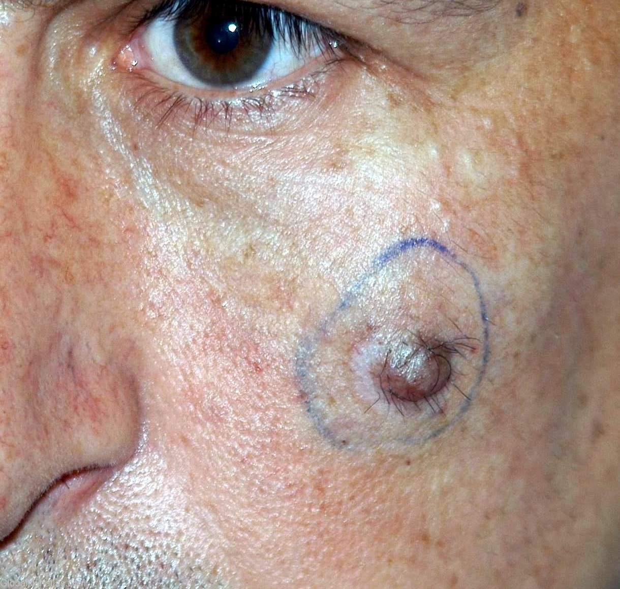
Description: Nodular lesion on the cheek
Morphology: Nodule,purple
Site: Cheek
Sex: M
Age: 30
Type: Clinical & Histology
Submitted By: Nasuhi Engin Aydin
Differential Diagnosis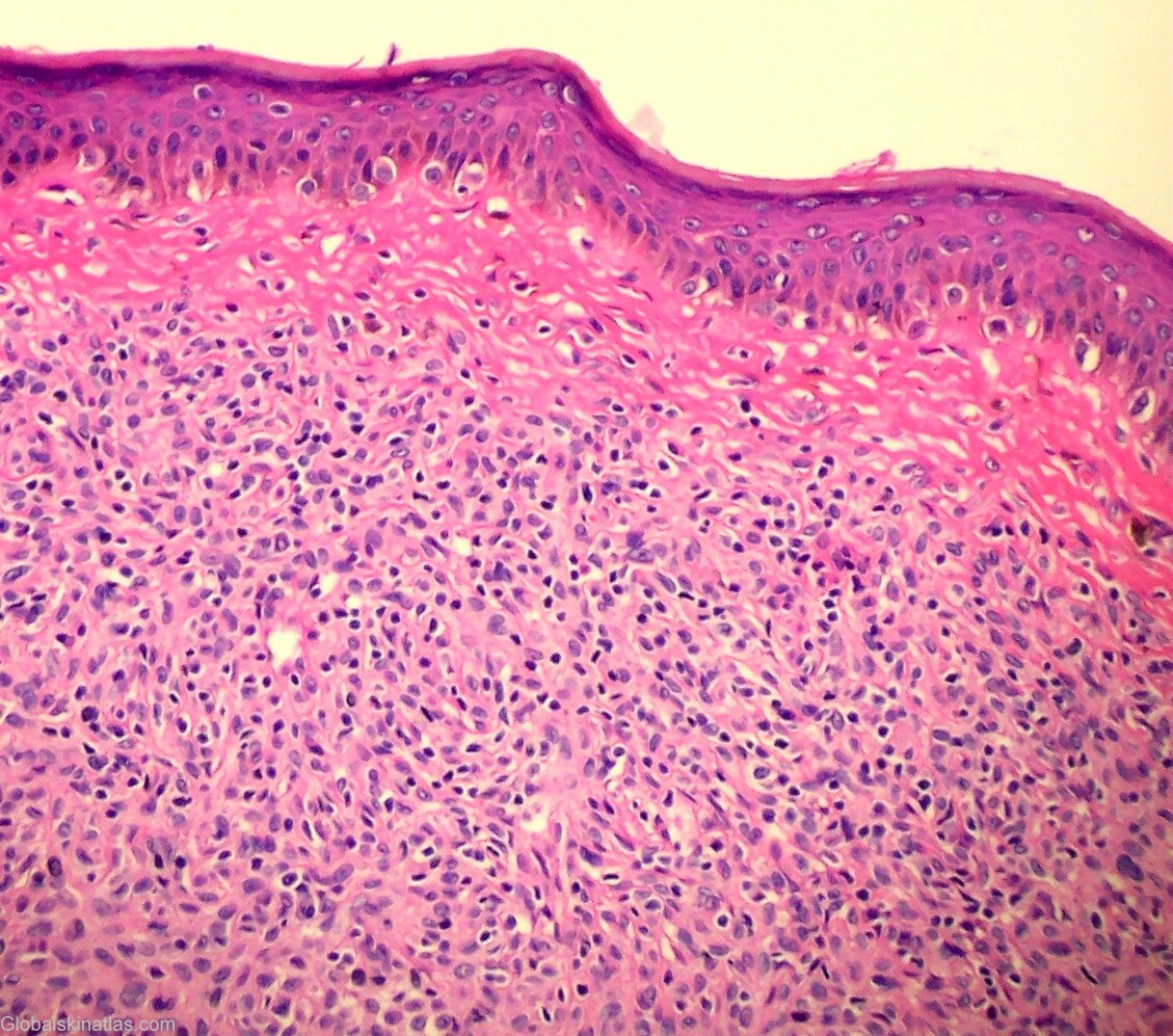
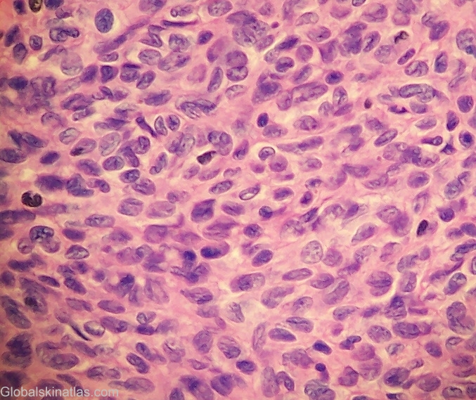
History: A paramedic noticed increase in size of a long-standing nodule on his cheek. Excision revealed a cellular dermal lesion with mitotic activity. However, there was no epidermal migration and the lesion was well demarcated. The final diagnosis ( the consensus of three pathologists from different countries) was cellular blue naevus. One of the consultants (a dermatologist from Graz, Austria) was more cautious and suggested close follow-up with a prefix "borderline malignancy". The patient is well without recurrence after a 2 year clinical surveillance.