
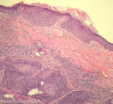
Diagnosis: Sebaceous carcinoma
Description: Low power view infiltrating carcinoma with in situ epidermal component
Morphology: Path,tumour
Site: Eyelids
Sex: F
Age: 55
Type: Histology
Submitted By: Nasuhi Engin Aydin
Differential Diagnosis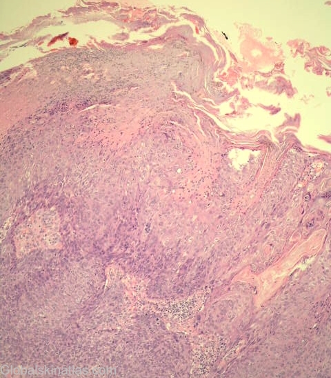
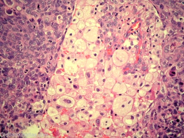
History: 55 year old woman with enlarging eyelid mass, about 2cm. Biopsy revealed sebaceous carcinoma which must be differentiated from squamous cell carcinoma (and sometimes from basal cell carcinoma) due to different clinical consequences. Sebaceous carcinoma of the periorbital region can be a diagnostic challenge for the clinican and pathologist. Immunohistochemistry may help in this regard ( see reference Sinard JH, Archives of Opthalmology, 117:776-83, 1999).