
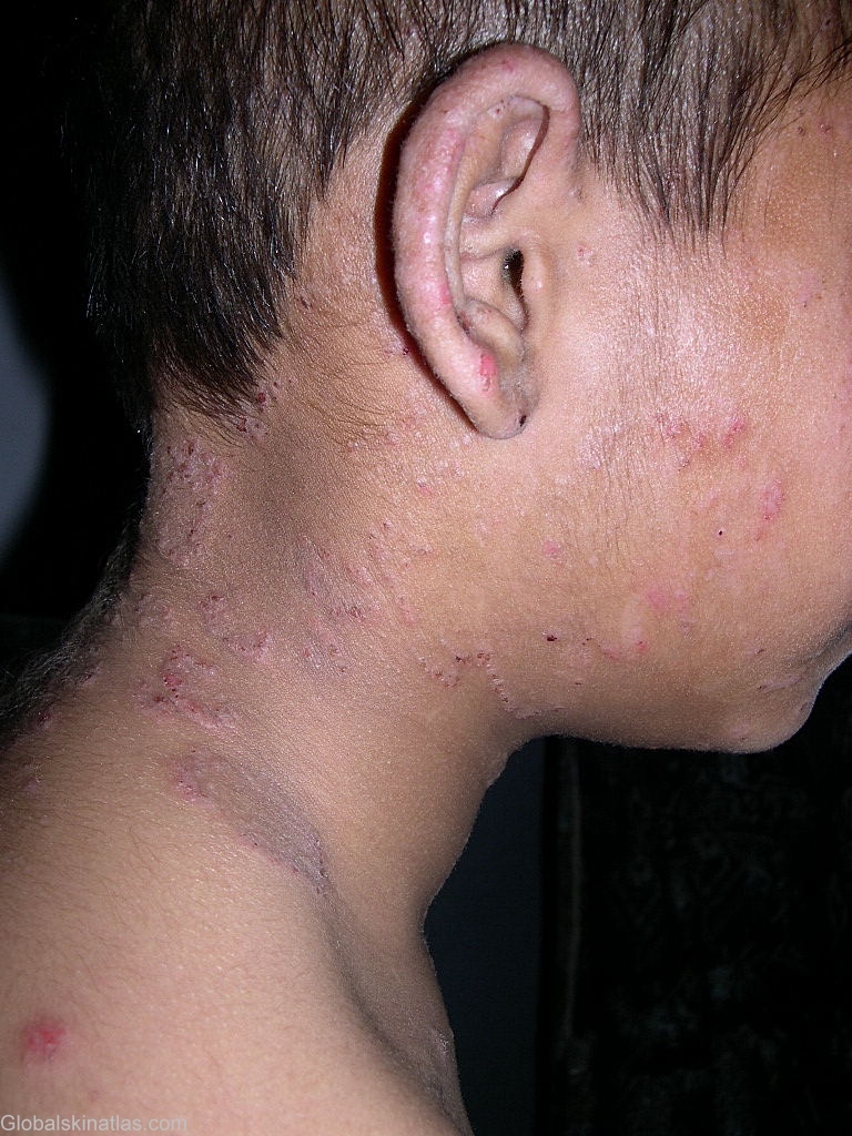
Diagnosis: Dermatophytosis
Description: Tinea corporis
Morphology: Red,scaly
Site: Neck side
Sex: M
Age: 12
Type: Clinical
Submitted By: Shahbaz Janjua
Differential Diagnosis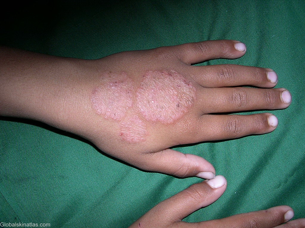
History:
Dermatophytosis or tinea is a fungal infection caused by dermatophytes. Several species of dermatophytes infect humans; these belong to the Epidermophyton, Microsporum, and Trichophyton genera. The infection may spread from person to person (anthropophilic), animal to person (zoophilic), or soil to person (geophilic). The most common of these organisms are Trichophyton rubrum, Trichophyton tonsurans, Trichophyton interdigitale and/or Trichophyton mentagrophytes, Microsporum canis, and Epidermophyton floccosum. Dermatophytes have the ability to invade keratinized tissue (eg, hair, nails, any area of the skin) but are restricted to the dead cornified layer of the epidermis. Humid or moist skin provides a very favorable environment for the establishment of fungal infection. Clinically, tinea infections are classified according to the body region involved. Tinea capitis is infection of scalp hair. Tinea corporis is infection of the trunk and extremities. Tinea manuum and tinea pedis is infection of palms, soles, and interdigital webs.Tinea cruris is infection of the groin. Tinea barbae is infection of the beard area and neck. Tinea faciale is infection of the face. Tinea unguium (onychomycosis) is infection of the nail.