
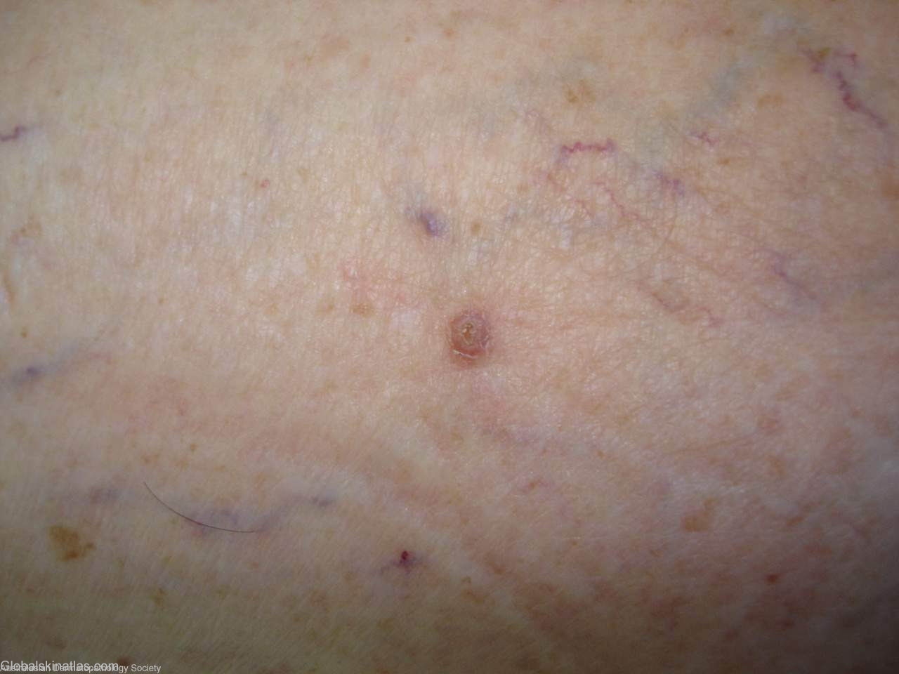
Diagnosis: Warty Dyskeratoma
Description: Small nodule on calf
Morphology: Nodule
Site: Leg
Sex: F
Age: 60
Type: Clinical & Histology
Submitted By: Ian McColl
Differential Diagnosis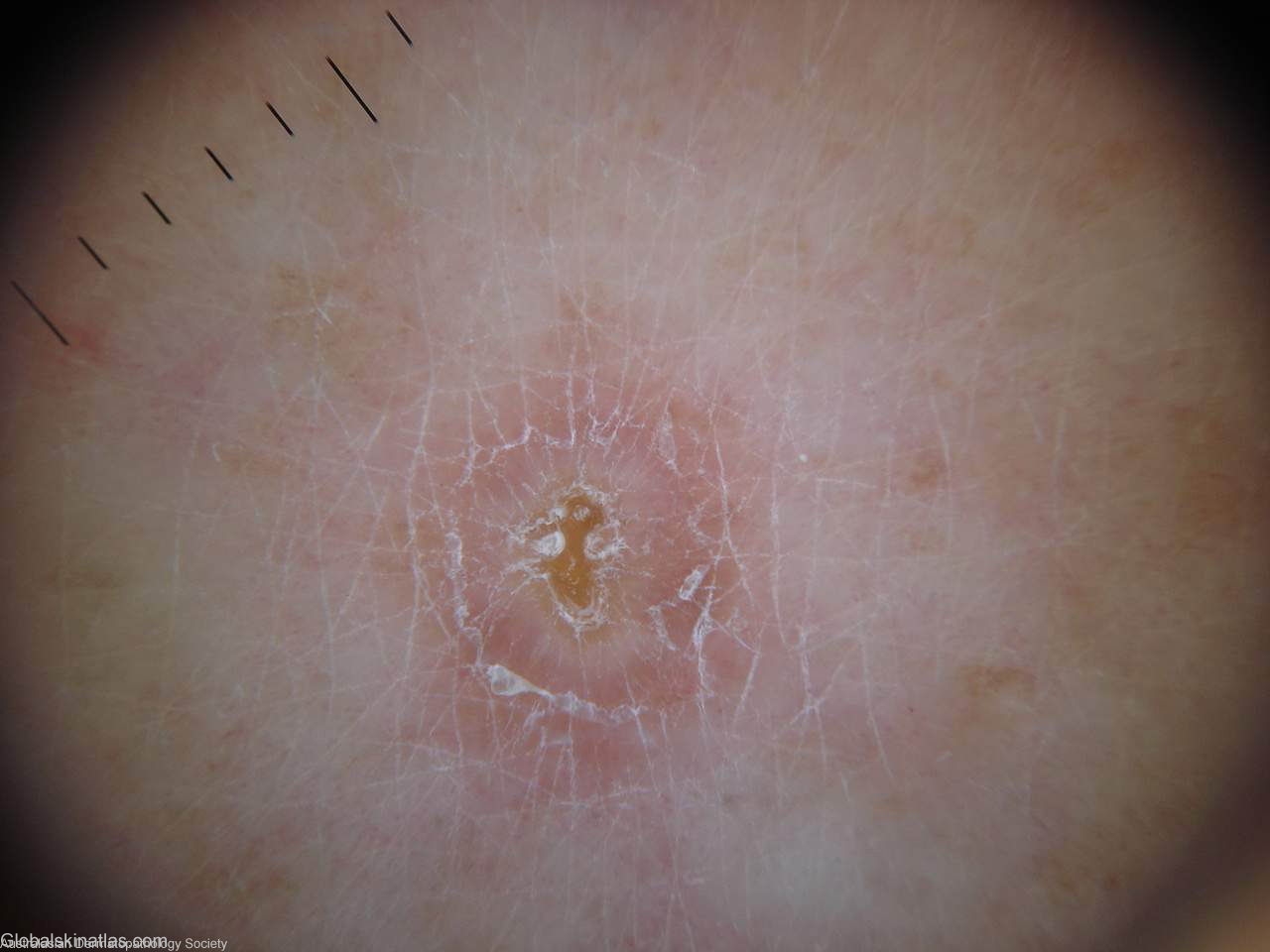
History:
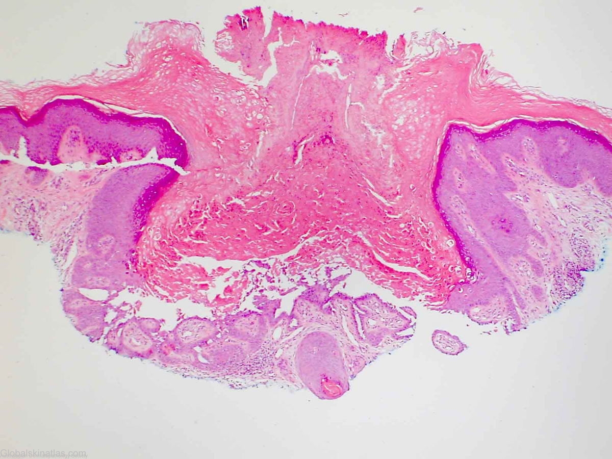
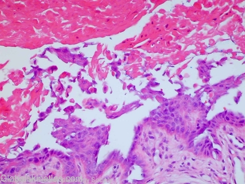
Keratotic lesion on the leg which has been irritable.Dermatoscope image suggests dermatofibroma but unlikely clinically.Sections show a warty dyskeratoma. There is a keratin plugged invagination in which there is acantholytic dyskeratosis and suprabasalar clefting. There is no evidence of malignancy.
Warty dyskeratomas are usually solitary lesions most commonly seen on the face.They have this pore like centre which may be extruded in the rare oral variant.