
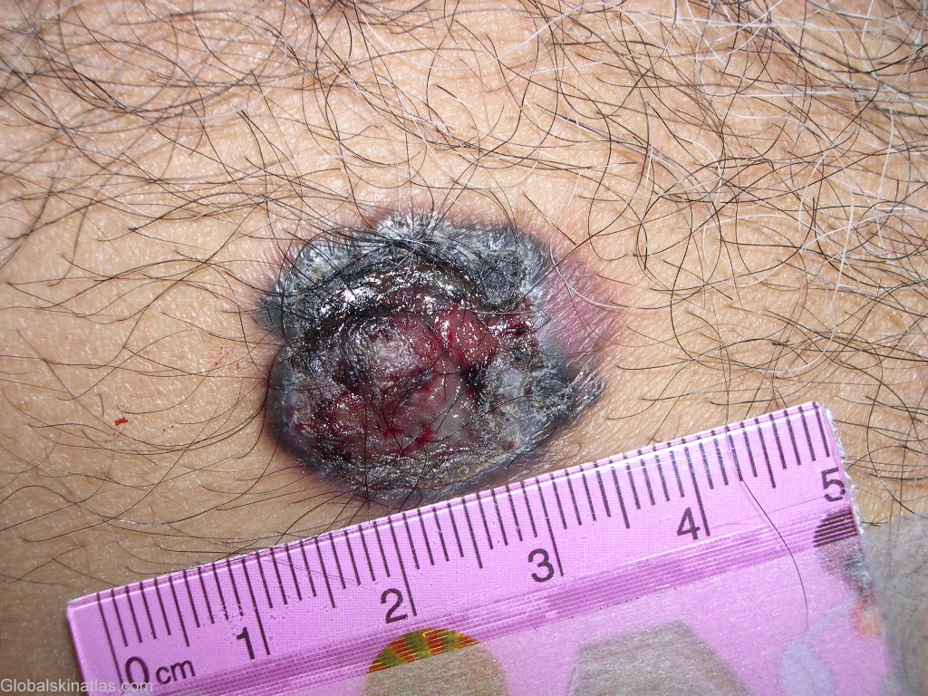
Diagnosis: Melanoma
Description: Ulcerated melanoma
Morphology: Ulcer
Site: Chest
Sex: M
Age: 70
Type: Clinical
Submitted By: Shahbaz Janjua
Differential DiagnosisHistory:
Microstaging is an integral part of the staging and clinical management of melanoma. Two methods have been used. The Breslow microstaging method measures the thickness of the lesion in millimeters using an ocular micrometer. The total vertical height of the melanoma is measured from the granular layer to the area of deepest penetration. The Clark method assesses the level of penetration into the various skin layers.
Clark Classification System of Microstaging
Level I:
Confined to the epidermis (in situ)
Level II:
Invasion into the papillary dermis
Level III:
Penetration to the papillary-reticular interface
Level IV:
Invasion into the reticular dermis
Level V:
Penetration into subcutaneous fat