
Diagnosis: Lymphoma Blastic NK cell type
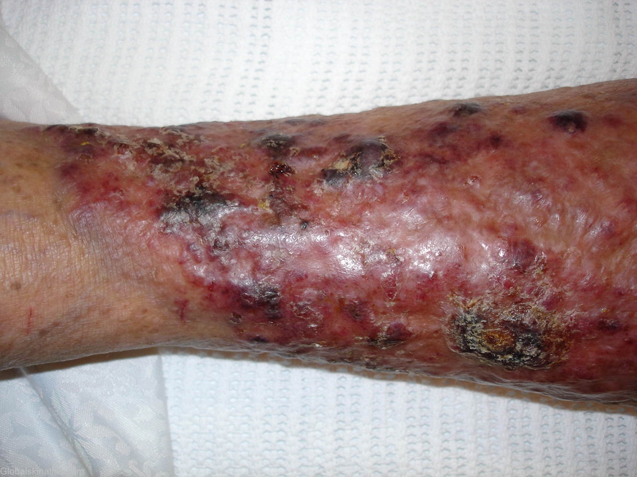
Description: Purple plaques and nodules
Morphology: Nodule,purple
Site: Leg
Sex: M
Age: 85
Type: Clinical & Histology
Submitted By: Ian McColl
Differential Diagnosis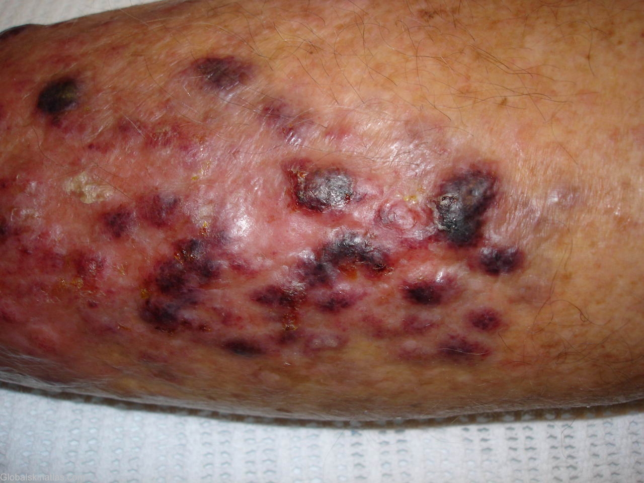

History: A 87 years old male presented with a history of a swelling on his lower leg that came up rapidly about 3 weeks before.This was diagnosed as a keratoacanthoma by a dermatologist who saw him but a week later he said he awoke one morning to find his lower leg swollen and covered in blue black nodules.He also developed pain and tenderness in the draining groin though he was not systemically ill.This is a classical case of blastic NK cell lymphoma which is a recently described entity. In a recent case series, plasmacytoid dendritic cell origin has been suggested for the blastic NK cell lymphoma
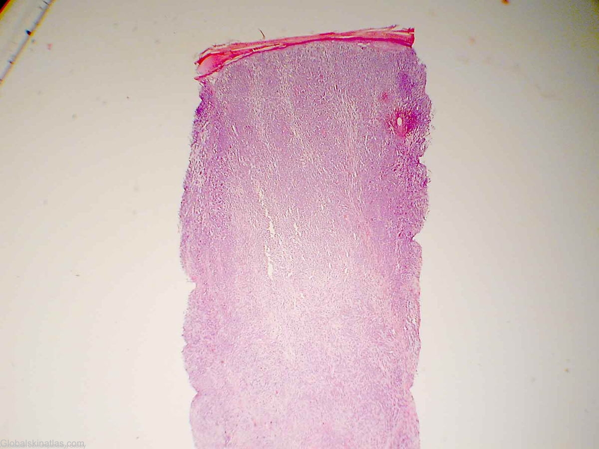
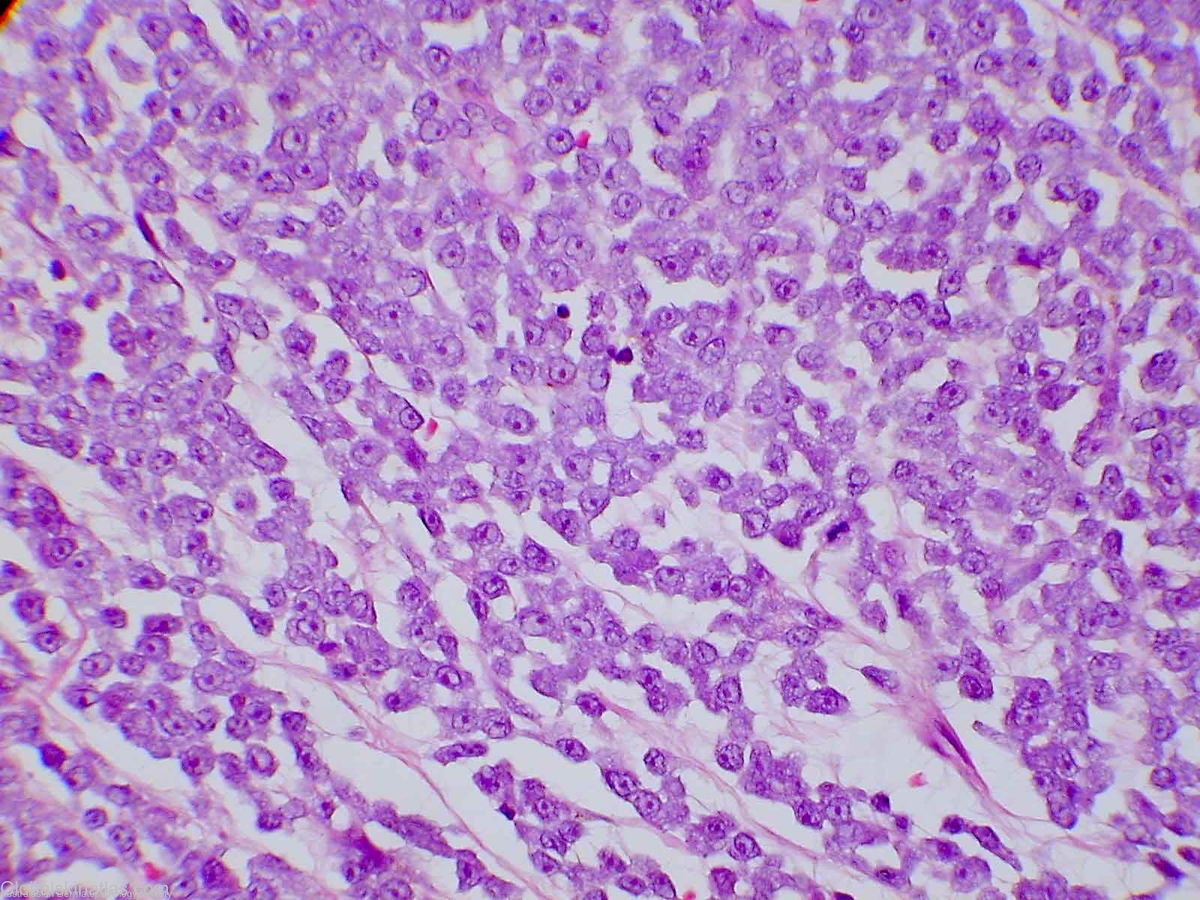
The pathology report was as follows The dermis is completely replaced and infiltrated by malignant tumour cells. The cells are medium in size and have small amounts of grey cytoplasm. The cell nuclei are moderately pleomorphic and contain fairly prominent amphophilic nucleoli. The cells completely fill the dermis forming sheets with some cell discohesion. The mitotic rate is high. In addition tumour cells invade the overlying epidermis in a focally pagetoid fashion. The tumour cells are positive for the pan leucocyte marker CD45. They are also positive for the CD43 and CD56 immunoperoxidase stains. The cells are negative for the CD3, CD5, CD10, CD20, CD30, CD38, CK20, myeloperoxidase, TTF-1, HMB45, S100 and AE1/AE3 immunoperoxidase stains. The morphology and immuno-profile of the tumour indicates that it is a blastic non-Hodgkins lymphoma. The positive CD56 and CD43 staining with concomitant negative CD3 and myeloperoxidase staining indicates a most likely diagnosis of blastic NK-cell lymphoma.