
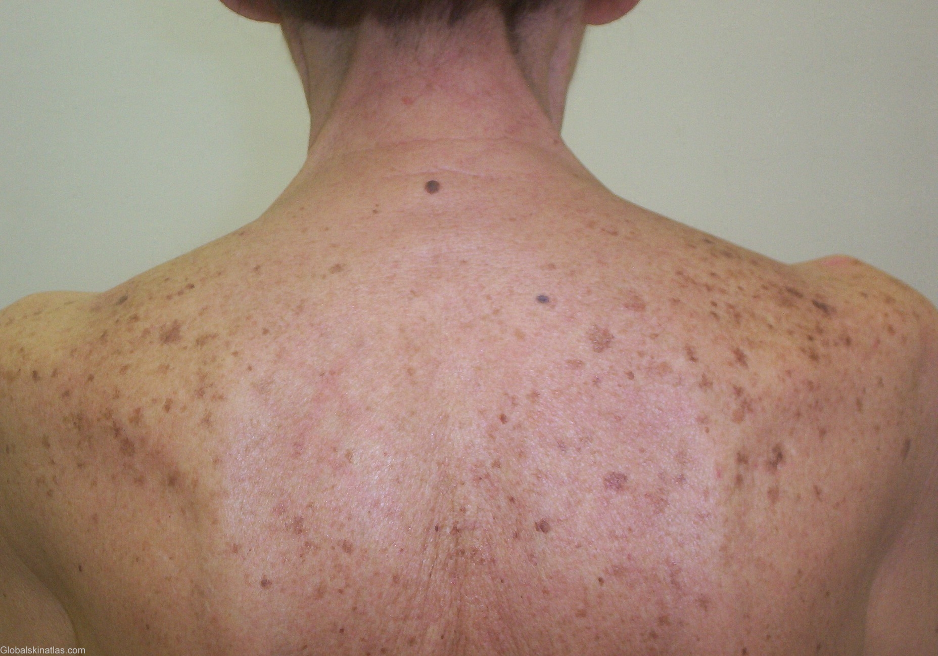
Diagnosis: Senile lentigines
Description: Multiple, symetrical, flat homogen brown lesions of the sun-exposed areas
Morphology: Macule brown
Site: Back
Sex: F
Age: 79
Type: Clinical
Submitted By: Mehravaran Mehrdad
Differential DiagnosisHistory: A 79-year-old female with multiple symetrical, homogeneous brown, flat macules and patches on her upper back and sholuders. Lentigo is a Latin word that means “lentil shaped” and has come to mean any lentil-shaped spot on the skin like a freckle. A lentigo is a well circumscribed brown to brown-black macule, ususally less than 1 cm in size. Lentigines occur in all skin types, and may be found on any cutaneous surface, including the palms, soles and mucous membranes and do not darken with sun exposure. Lentigines can be localized or generalized and must be distinguished from freckles /ephelides/. The histopathology of lentigines shows elongated rete ridges, increased numbers of basal melanocytes, and increased basal melanisation. In contrast, freckles result from hypermelanisation of basal melanocytes without a concomitant increase in their number. Several types of lentigines are recognized, including lentigo simplex, generalized lentiginosis, senile lentigines, nevus spilus, and lentigines induced by PUVA. Senile lentigines or “liver spots” are brown macules which appear late in adult life on chronically sun-exposed skin. These lesions are present in 90% of white people over 70.