
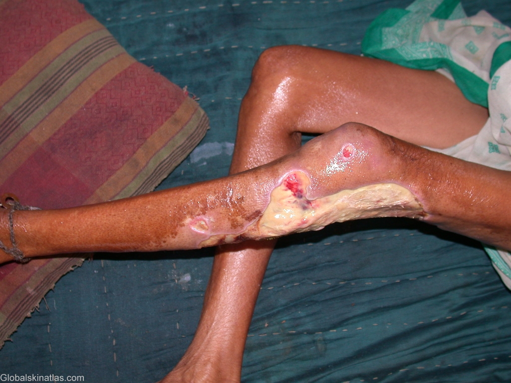
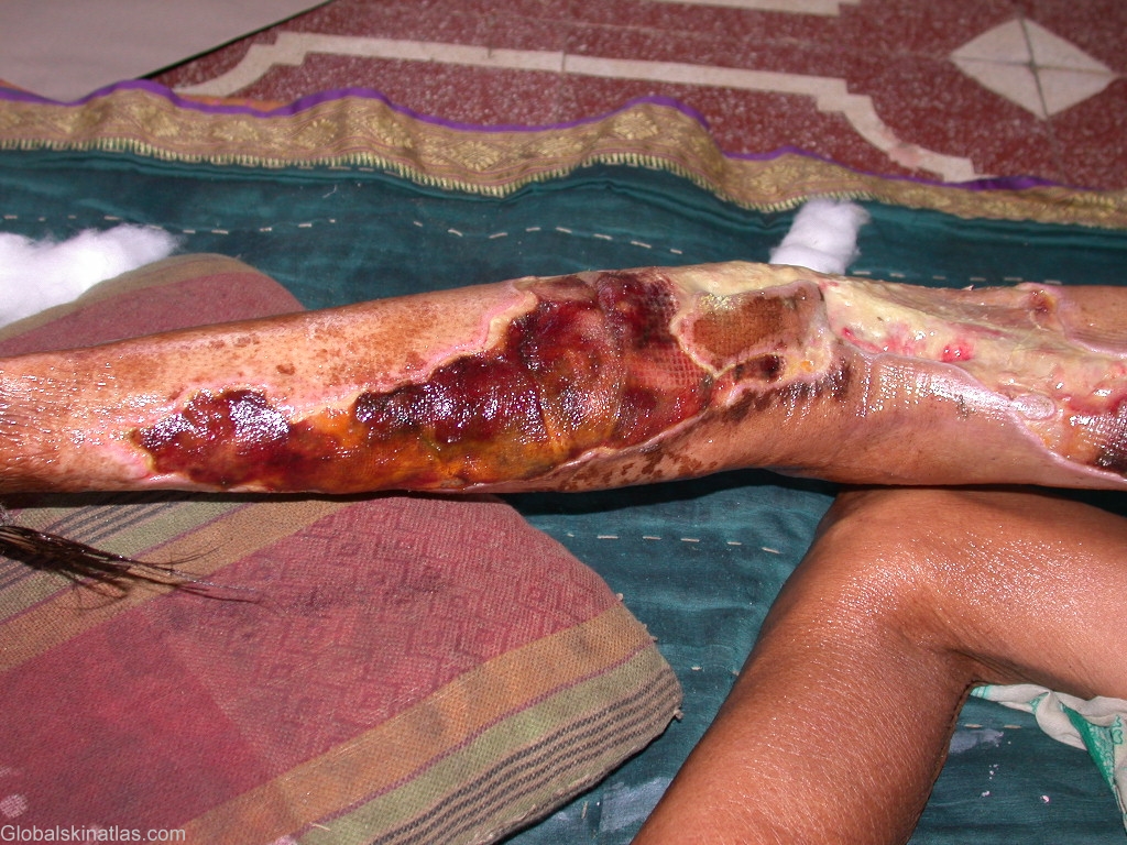
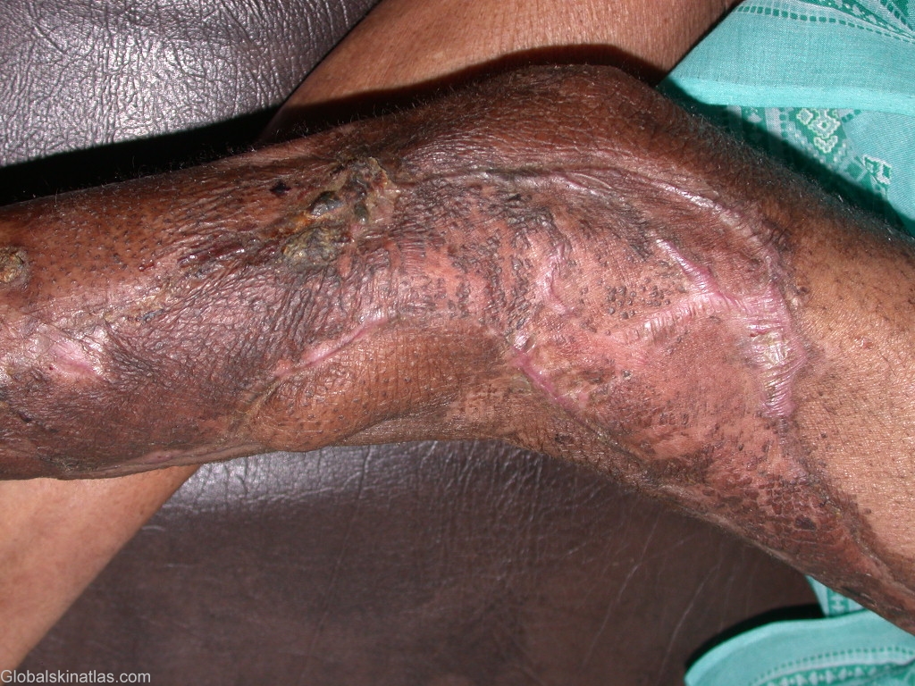
Diagnosis: Necrotizing Fasciitis
Description: Skin necrosis,Deep widespread ulcers
Morphology: Ulcer
Site: Leg
Sex: F
Age: 55
Type: Clinical
Submitted By: Kiran Nabar
Differential Diagnosis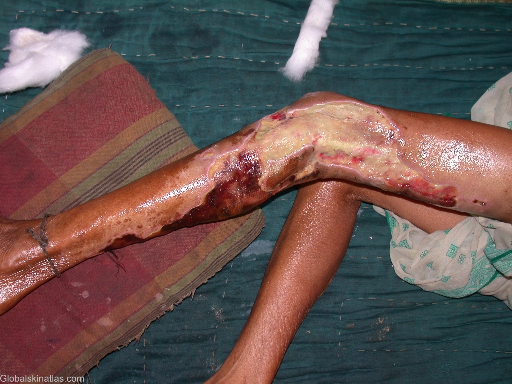
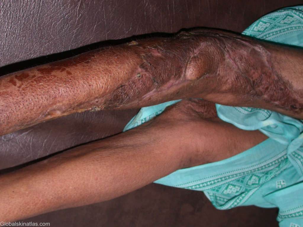
History:
This 55 yr old lady had a history of a thorn prick injury on the left knee with a small 3mm diameter penetrating wound. After 8 days she had a fever with chills, followed by painful erythema and swelling of the left foot below the knee. Within the next 2 days a few blisters occurred on the affected skin which then became necrosed and sloughed off leaving behind deep widespread ulcers with undermined edges and subcutaneous fat in the floor. Debridement of necrosed skin was done under general anaesthesia, i.v. antibiotics were started and after 2 weeks split thickness skin grafting was done
Necrotising Fasciitis is an uncommon form of pyoderma .It is caused by Group A beta Hemolytic Streptococci .Bacteria enter deep in the skin after some penetrating injury. Infection travels at the fascial level .Blood vessels passing through the fascia and supplying the overlying skin are also affected leading to necrosis of the skin.