
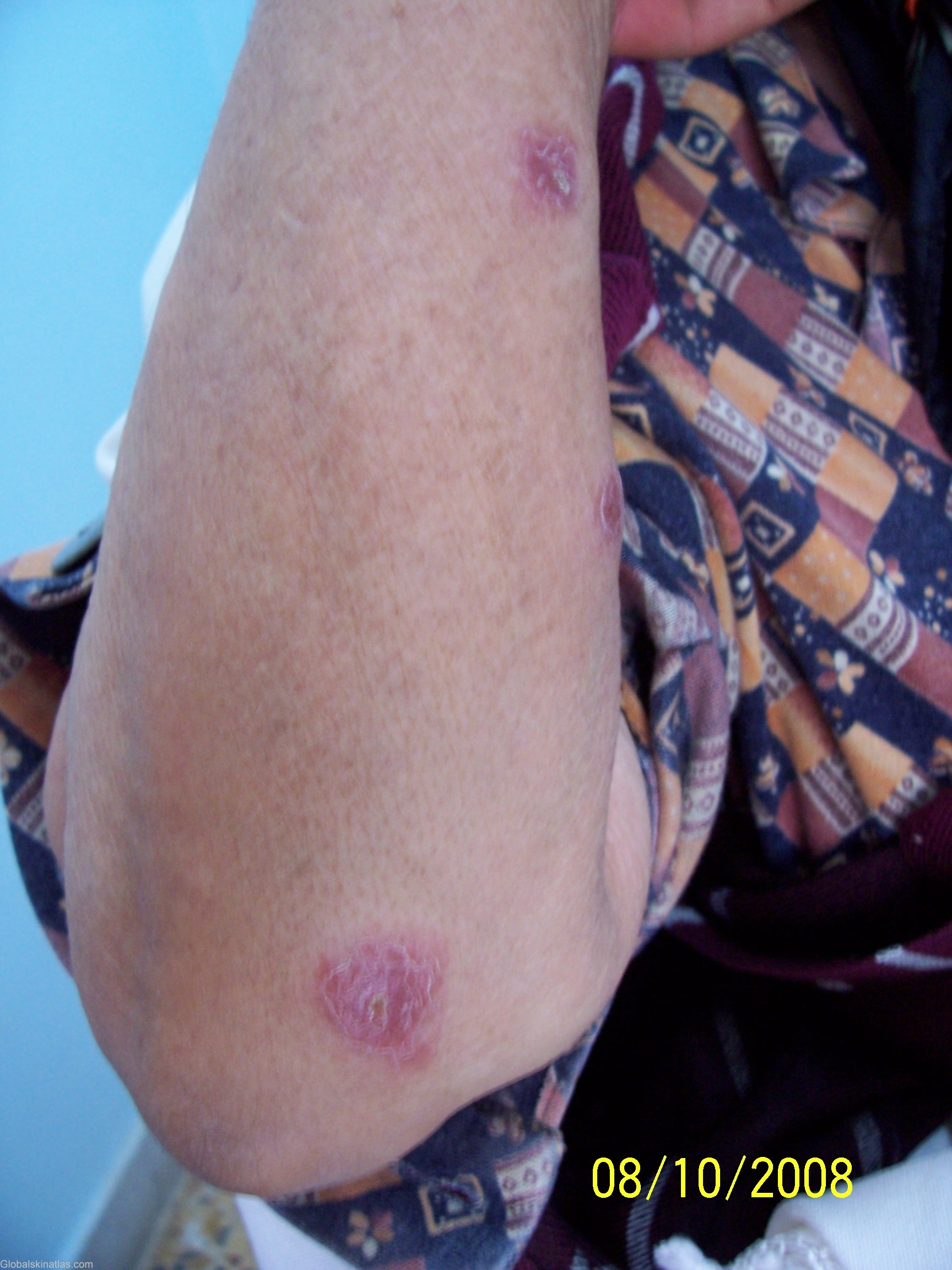
Diagnosis: Leishmaniasis
Description: right forearm showing 2 papules
Morphology: Ulcer
Site: Arm,forearm
Sex: F
Age: 60
Type: Clinical
Submitted By: Ebtisam Elghblawi
Differential Diagnosis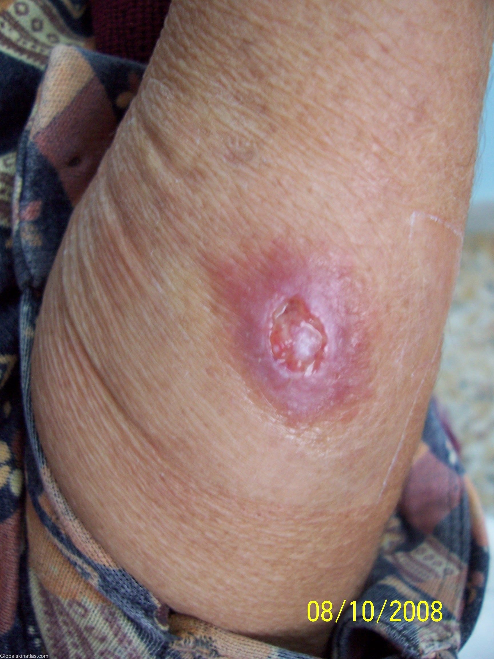
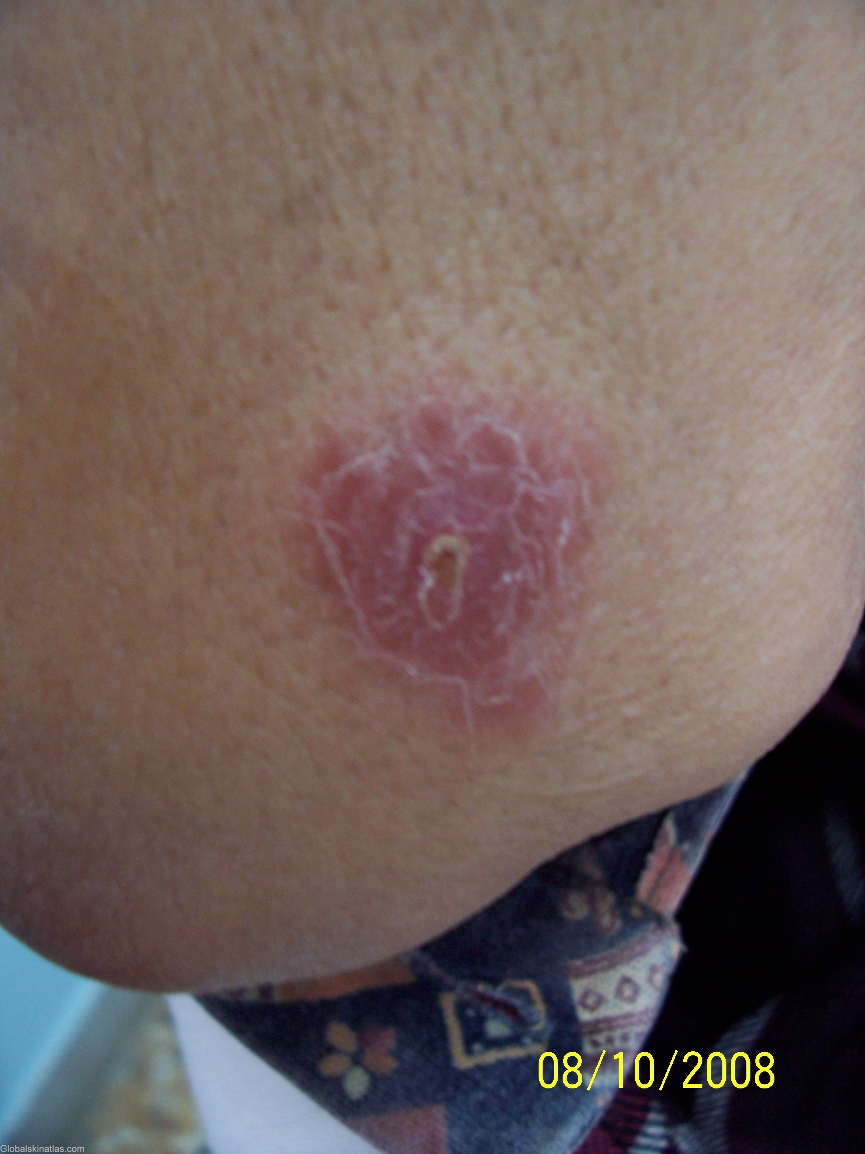
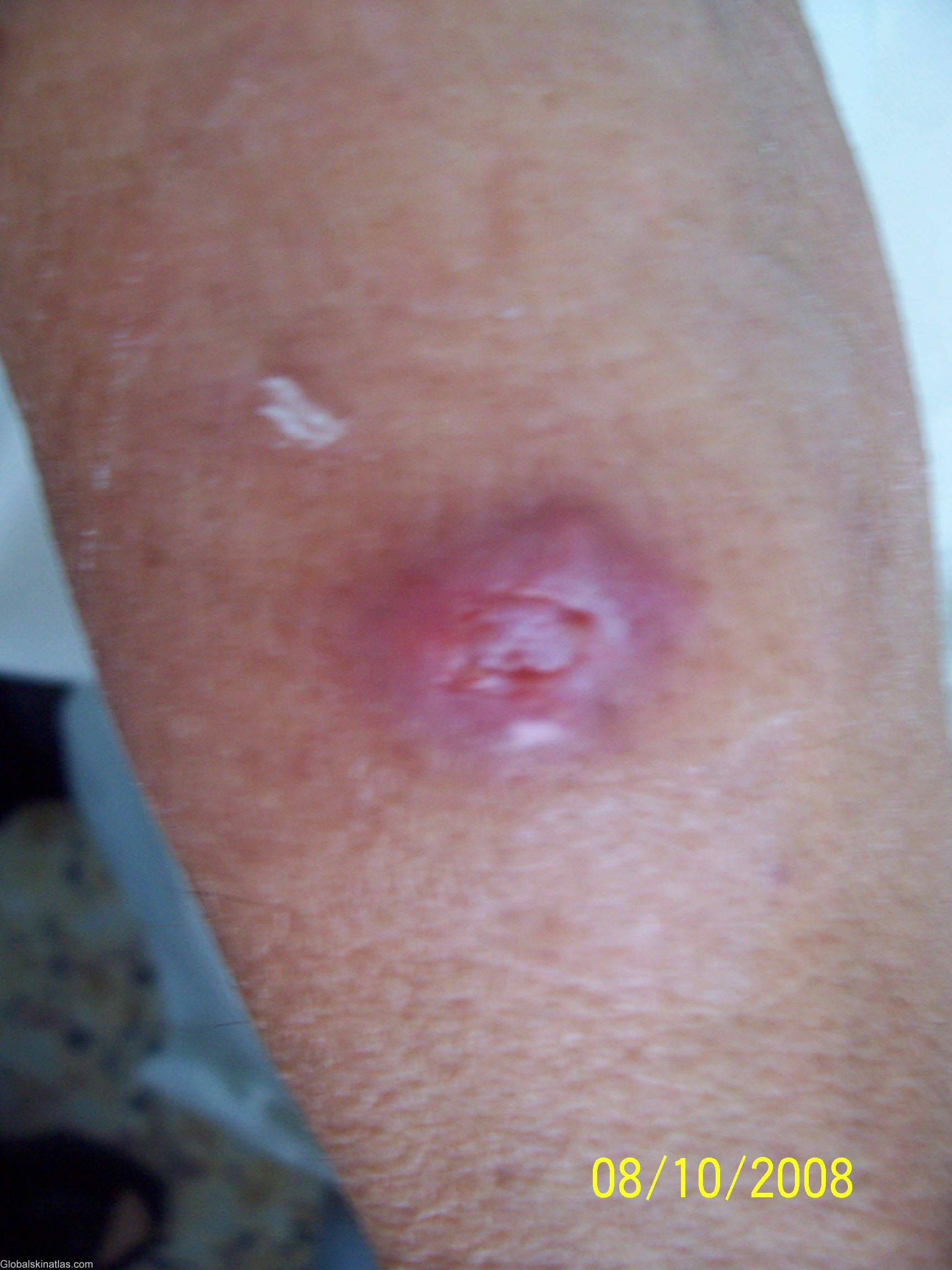

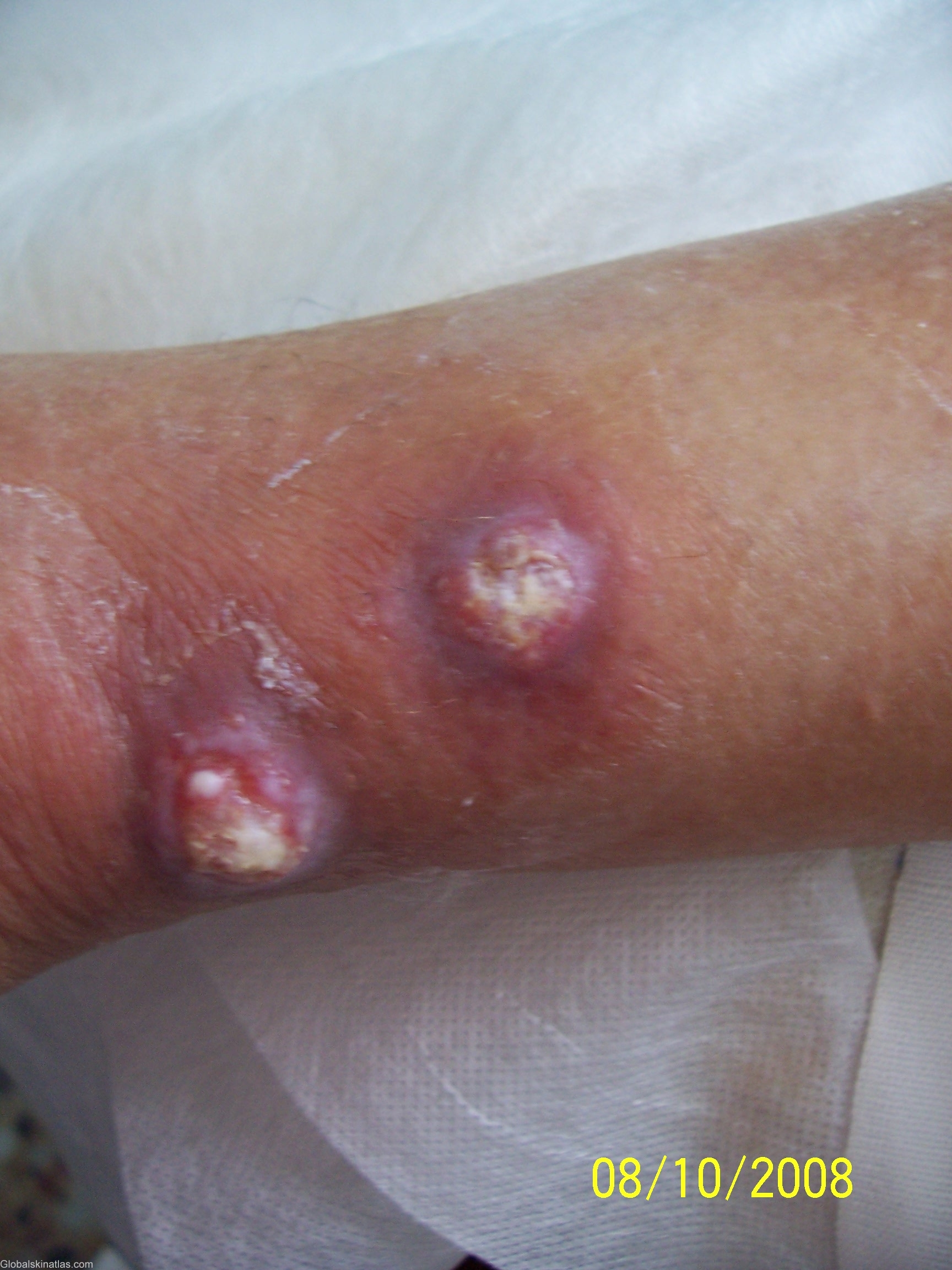
History: An old lady 60 years old presented with a history of two months papules and ulcers all over her extremities. Some are still papules and some open up into ulcers. No other member of her family affected. She is diabetic on insulin since 8 years back. Leishmania is a protozoan infection that is responsible for three primary diseases; Systemic or Visceral leishmaniasis, Cutaneous leishmaniasis, and lastly Mucocutaneous infection. Cutaneous leishmaniasis, is the most common form of leishmaniasis in which the epidermis is the primary site of infection. It can either macule or papule erythematous (small raised skin lesions), or expanding skin ulcer. There are two different species that cause this; either L. tropica or L. major. They both have the same symptoms and life cycles but differ in their geographic distribution. Both species can produce large numbers of skin ulcers on the exposed parts of the body, such as the face, arms and legs, causing serious disability and leaving the patient permanently scarred. The infection site is usually localized to the site in which the sand fly bite occurs.