
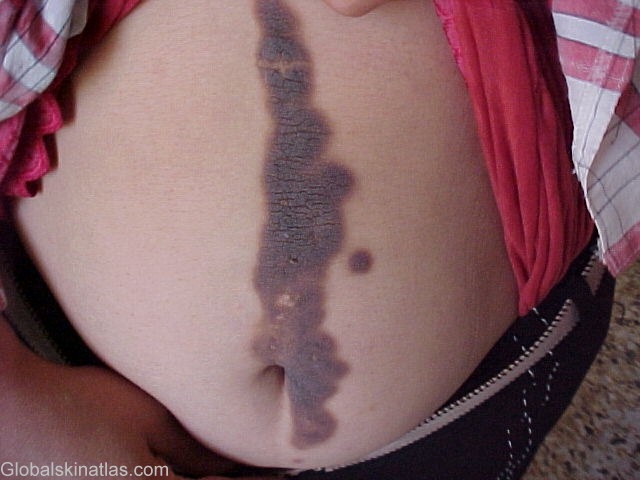
Diagnosis: Epidermal nevus
Description: well-demarcated, brown, papillomatous plaques
Morphology: Plaque
Site: Abdomen
Sex: F
Age: 33
Type: Clinical
Submitted By: Ebtisam Elghblawi
Differential DiagnosisHistory: Epidermal nevi are congenital hamartomas of embryonal ectodermal origin classified on the basis of their main component; the component may be sebaceous, apocrine, eccrine, follicular, or keratinocytic. Verrucous epidermal nevus is well-demarcated, skin coloured to brown, papillomatous plaques. Most lesions are present at birth or develop during early infancy; they enlarge slowly during childhood and generally reaching a stable size at adolescence. Lesions may be localized or diffuse. Linear configurations are common, especially on the limbs, and may follow skin tension lines, or Blaschko's lines.