
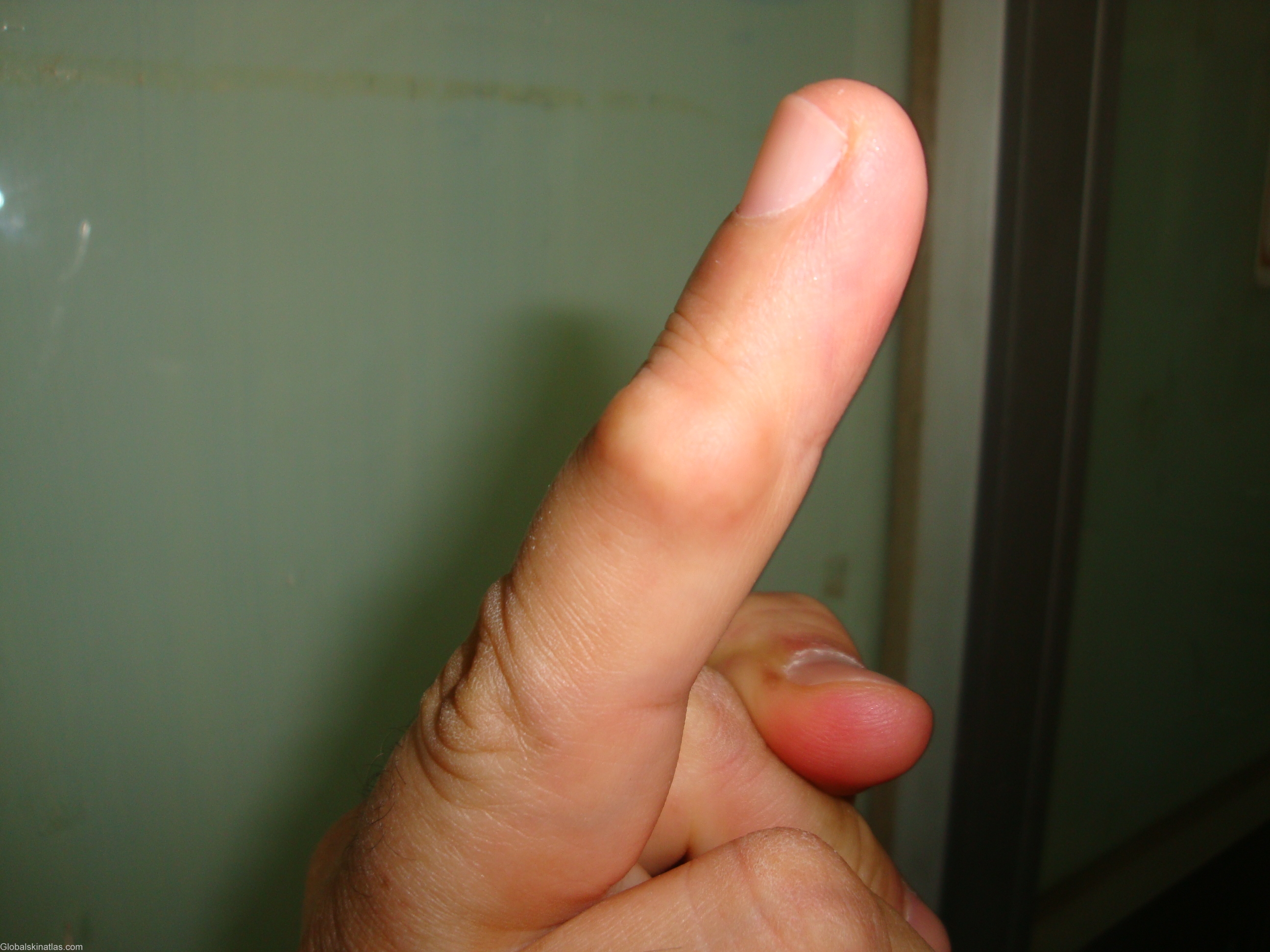
Diagnosis: Giant-cell tumour of tendon sheath
Description: Ruberry firm lobulated nodule
Morphology: Nodule
Site: Finger
Sex: M
Age: 39
Type: Clinical
Submitted By: Nameer Al-Sudany
Differential Diagnosis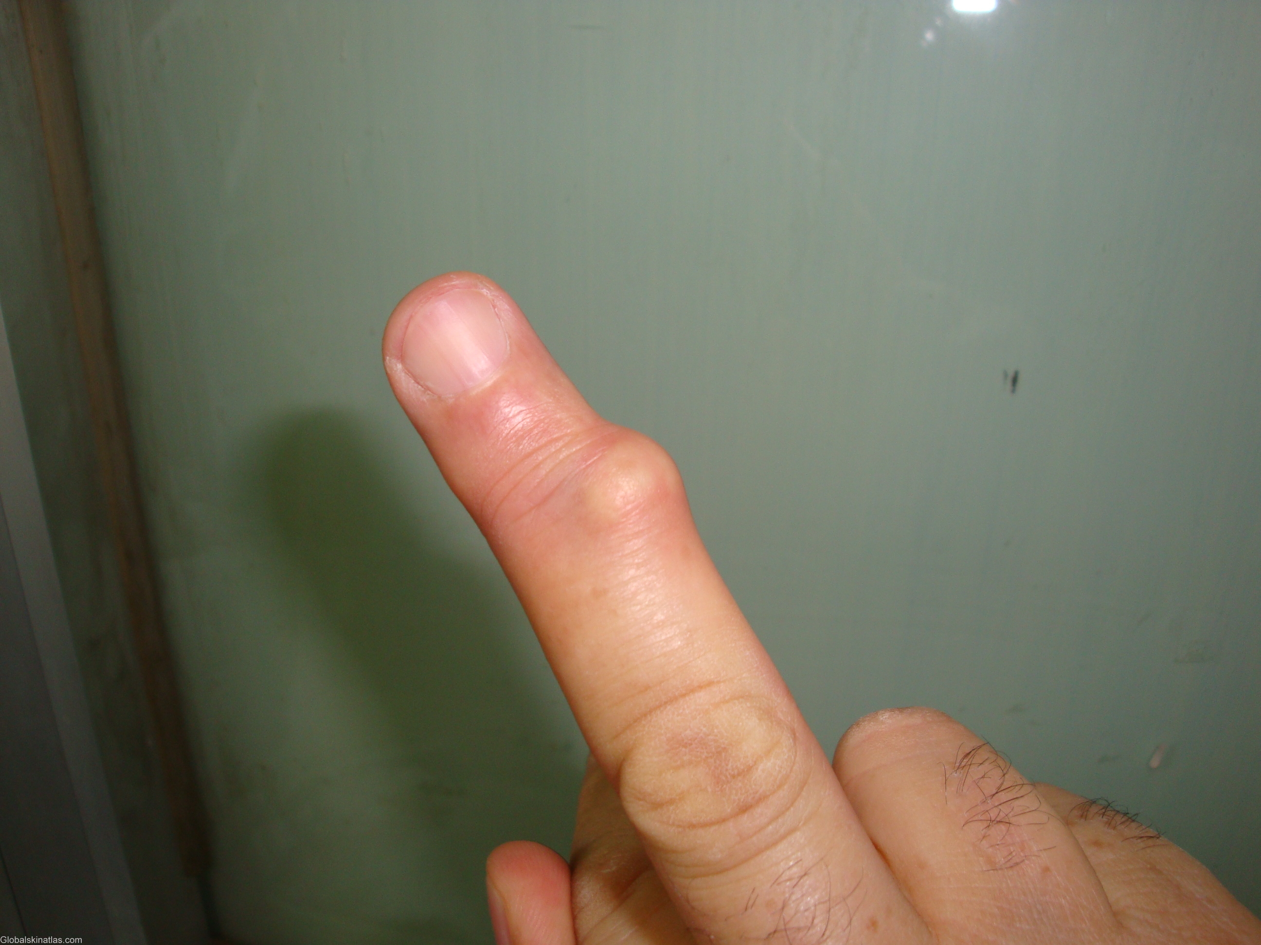
History:
Giant-cell tumour of tendon sheath
It is also called giant-cell synovioma or localized nodular tenosynovitis. It is a firm, dermal or subcutaneous nodule, composed of scattered multinucleated giant cells set on a background of proliferating spindle cells in a collagenous matrix. It usually occurs on the fingers. The cause is unknown and usually presents in mid-life as a single, firm, rubbery, subcutaneous mass on the finger. The nodule is usually asymptomatic, but in occasional cases there may be pain, numbness or stiffness of the digit. There is a strong association with osteoarthritis. The lesions do not regress, and they can persist for more than 20 years.