
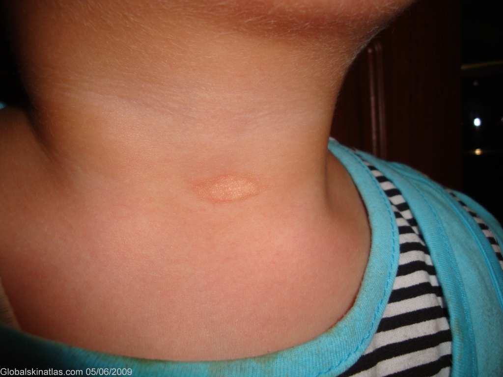
Diagnosis: Anetoderma
Description: Congenital, oval, atrophic, white patch on the front of the neck.
Morphology: Atrophy
Site: Neck front
Sex: F
Age: 2
Type: Clinical
Submitted By: Nameer Al-Sudany
Differential Diagnosis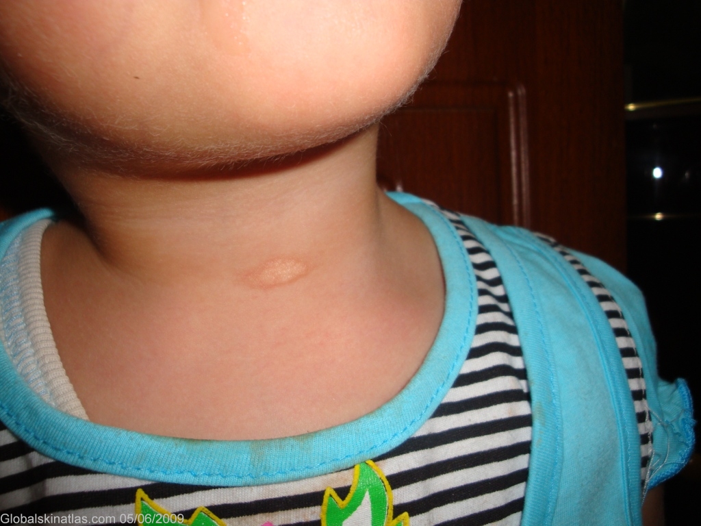
History:
Anetoderma (macular atrophy) is characterized by atrophic patches located mainly on the upper trunk. The skin of the patches is thin and blue-white and bulges slightly. The lesions may give the palpating finger the same sensation as a hernial orifice. In many patients, new lesions continue to appear over a period of several years. Rare congenital and familial cases have been reported. This child has an oval atrophic hypopigmented solitary patch with slightly hyperpigmented border on the front of her neck. It is about 3 cm x 1.5 cm diameters and has been noted by the mother since birth with constant size. There is no similar family history and it wasn’t preceded by any skin disease.